Growth patterns in the developing brain detected by using continuum mechanical tensor maps
- Select a language for the TTS:
- UK English Female
- UK English Male
- US English Female
- US English Male
- Australian Female
- Australian Male
- Language selected: (auto detect) - EN
Play all audios:
ABSTRACT The dynamic nature of growth and degenerative disease processes requires the design of sensitive strategies to detect, track and quantify structural change in the brain in its full
spatial and temporal complexity1. Although volumes of brain substructures are known to change during development2, detailed maps of these dynamic growth processes have been unavailable. Here
we report the creation of spatially complex, four-dimensional quantitative maps of growth patterns in the developing human brain, detected using a tensor mapping strategy with greater
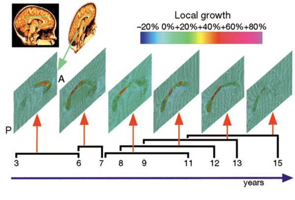
spatial detail and sensitivity than previously obtainable. By repeatedly scanning children (aged 3–15 years) across time spans of up to four years, a rostro-caudal wave of growth was
detected at the corpus callosum, a fibre system that relays information between brain hemispheres. Peak growth rates, in fibres innervating association and language cortices, were attenuated
after puberty, and contrasted sharply with a severe, spatially localized loss of subcortical grey matter. Conversely, at ages 3–6 years, the fastest growth rates occurred in frontal
networks that regulate the planning of new actions. Local rates, profiles, and principal directions of growth were visualized in each individual child. Access through your institution Buy or
subscribe This is a preview of subscription content, access via your institution ACCESS OPTIONS Access through your institution Subscribe to this journal Receive 51 print issues and online
access $199.00 per year only $3.90 per issue Learn more Buy this article * Purchase on SpringerLink * Instant access to full article PDF Buy now Prices may be subject to local taxes which
are calculated during checkout ADDITIONAL ACCESS OPTIONS: * Log in * Learn about institutional subscriptions * Read our FAQs * Contact customer support SIMILAR CONTENT BEING VIEWED BY OTHERS
MULTIFACETED ATLASES OF THE HUMAN BRAIN IN ITS INFANCY Article Open access 30 December 2022 REGIONAL PATTERNS OF HUMAN CORTEX DEVELOPMENT CORRELATE WITH UNDERLYING NEUROBIOLOGY Article Open
access 12 September 2024 HUMAN LIFESPAN CHANGES IN THE BRAIN’S FUNCTIONAL CONNECTOME Article 03 April 2025 REFERENCES * Fox, N. C., Freeborough, P. A. & Rossor, M. N. Visualisation and
quantification of rates of atrophy in Alzheimer's disease. _Lancet_ 348, 94– 97 (1996). Article CAS Google Scholar * Giedd, J. N. _ et al_. Quantitative magnetic resonance imaging of
human brain development: ages 4–18. _Cereb. Cortex_ 6, 551– 560 (1996). Article CAS Google Scholar * Yakovlev, P. I. & Lecours, A. R. in _Regional Development of the Brain in Early
Life_ (ed. Minkowski, A.) 3– 70 (Davis, Philadelphia, 1967). Google Scholar * Sowell, E. R., Thompson, P. M., Holmes, C. J., Jernigan, T. L. & Toga, A. W. _In vivo_ evidence for
post-adolescent brain maturation frontal and striatal regions. _Nature Neurosci._ 2, 859– 861 (1999). Article CAS Google Scholar * Chugani, H. T., Phelps, M. E. & Mazziotta, J. C.
Positron emission tomography study of human brain functional development. _Ann. Neurol._ 22, 487–497 (1987). Article CAS Google Scholar * Grimshaw, G. M., Adelstein, A., Bryden, M. P.
& MacKinnon, G. E. First-language acquisition in adolescence: evidence for a critical period for verbal language development. _Brain Lang._ 63, 237–255 (1998). Article CAS Google
Scholar * Thompson, P. M. _ et al_. Cortical variability and asymmetry in normal aging and Alzheimer's disease. _Cereb. Cortex_ 8, 492– 509 (1998). Article CAS Google Scholar *
Zijdenbos, A. P. & Dawant, B. M. Brain segmentation and white matter lesion detection in MR images. _Crit. Rev. Biomed. Eng._ 22, 401–465 ( 1994). CAS PubMed Google Scholar * Woods,
R. P., Cherry, S. R. & Mazziotta, J. C. Rapid automated algorithm for aligning and reslicing PET images. _J. Comp. Assist. Tomogr._ 16, 620–633 (1992). Article CAS Google Scholar *
Freeborough, P. A., Woods, R. P. & Fox, N. C. Accurate registration of serial 3D MR brain images and its application to visualizing change in neurodegenerative disorders. _ J. Comp.
Assist. Tomogr._ 20, 1012– 1022 (1996). Article CAS Google Scholar * MacDonald, D., Avis, D. & Evans, A. C. in _Proc. SPIE Conf. Visualization in Biomedical Computing _ (ed. Robb, R.
A.) 2359, 160–169 (1994). Google Scholar * Thompson, P. M. & Toga, A. W. A surface-based technique for warping 3-dimensional images of the brain. _IEEE Trans. Med. Imag._ 15, 471–489 (
1996). Article Google Scholar * Thompson, P. M. & Toga, A. W. Detection, visualization and animation of abnormal anatomic structure with a deformable probabilistic brain atlas based on
random vector field transformations. _Med. Image Anal._ 1, 271–294 ( 1997). Article CAS Google Scholar * Thompson, P. M. & Toga, A. W. in _Brain Warping _ (ed. Toga, A. W.) 311–336
(Academic, San Diego, 1998). Google Scholar * Thompson, P. M., Schwartz, C., Lin, R. T., Khan, A. A. & Toga, A. W. 3D statistical analysis of sulcal variability in the human brain. _J.
Neurosci._ 16, 4261– 4274 (1996). Article CAS Google Scholar * Thompson, P. M. _ et al_. Detection and mapping of abnormal brain structure with a probabilistic atlas of cortical surfaces.
_J. Comp. Assist. Tomogr._ 21, 567–581 (1997). Article CAS Google Scholar * Davatzikos, C. Spatial normalization of 3D brain images using deformable models. _ J. Comp. Assist. Tomogr._
20, 656–665 (1996). Article CAS Google Scholar * Miller, M. I. & Grenander, U. Computational anatomy: an emerging discipline. _Q. Appl. Math._ 56, 617– 694 (1998). Article MathSciNet
Google Scholar Download references ACKNOWLEDGEMENTS We thank E. Sowell, M. Mega and J. Mazziotta for their advice and support. P.M.T. was supported by the Howard Hughes Medical Institute,
the US Information Agency, and the US–UK Fulbright Commission. Additional research support was provided by a Human Brain Project grant to the International Consortium for Brain Mapping,
funded jointly by NIMH and NIDA, by National Institutes of Health intramural funding (J.N.G.), and by the National Library of Medicine, National Science Foundation, and the NCRR. AUTHOR
INFORMATION AUTHORS AND AFFILIATIONS * Department of Neurology, Division of Brain Mapping, Laboratory of Neuro Imaging, UCLA School of Medicine, 710 Westwood Plaza, Los Angeles, 90095-1769,
California, USA Paul M. Thompson, Roger P. Woods & Arthur W. Toga * Child Psychiatry Branch, National Institute of Mental Health, NIH, 10 Center Drive, Bethesda, MSC 1600, 20982-1600,
Maryland, USA Jay N. Giedd * Montreal Neurological Institute, McGill University, 3801 University Street, Montreal , H3A 2B4, Québec, Canada David MacDonald & Alan C. Evans Authors * Paul
M. Thompson View author publications You can also search for this author inPubMed Google Scholar * Jay N. Giedd View author publications You can also search for this author inPubMed Google
Scholar * Roger P. Woods View author publications You can also search for this author inPubMed Google Scholar * David MacDonald View author publications You can also search for this author
inPubMed Google Scholar * Alan C. Evans View author publications You can also search for this author inPubMed Google Scholar * Arthur W. Toga View author publications You can also search for
this author inPubMed Google Scholar CORRESPONDING AUTHOR Correspondence to Arthur W. Toga. RIGHTS AND PERMISSIONS Reprints and permissions ABOUT THIS ARTICLE CITE THIS ARTICLE Thompson, P.,
Giedd, J., Woods, R. _et al._ Growth patterns in the developing brain detected by using continuum mechanical tensor maps. _Nature_ 404, 190–193 (2000). https://doi.org/10.1038/35004593
Download citation * Received: 27 August 1999 * Accepted: 21 January 2000 * Issue Date: 09 March 2000 * DOI: https://doi.org/10.1038/35004593 SHARE THIS ARTICLE Anyone you share the following
link with will be able to read this content: Get shareable link Sorry, a shareable link is not currently available for this article. Copy to clipboard Provided by the Springer Nature
SharedIt content-sharing initiative