Publisher correction: juvenile depletion of microglia reduces orientation but not high spatial frequency selectivity in mouse v1
- Select a language for the TTS:
- UK English Female
- UK English Male
- US English Female
- US English Male
- Australian Female
- Australian Male
- Language selected: (auto detect) - EN
Play all audios:
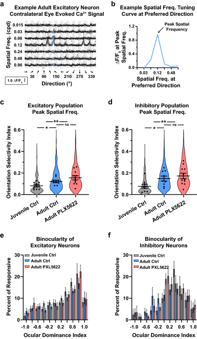
Correction to: _Scientific reports_ https://doi.org/10.1038/s41598-022-15503-0, published online 27 July 2022 The original version of this Article contained errors in Figure 3c and d, where
the y-axis labels were incorrectly given. As a result, Orientation Selectivity Index now reads: Spatial Frequency (cpd) Orientation Selectivity Index now reads: Spatial Frequency (cpd) The
original Figure 3 and accompanying legend appear below. The original Article has been corrected. AUTHOR INFORMATION AUTHORS AND AFFILIATIONS * Department of Pathology, Children’s Hospital
Boston, Boston, MA, 02115, USA Dario X. Figueroa Velez * Department of Neurobiology and Behavior, University of California, Irvine, CA, 92697, USA Miguel Arreola, Carey Y. L. Huh, Kim Green
& Sunil P. Gandhi * Center for the Neurobiology of Learning and Memory, University of California, Irvine, Irvine, CA, 92697, USA Kim Green & Sunil P. Gandhi Authors * Dario X.
Figueroa Velez View author publications You can also search for this author inPubMed Google Scholar * Miguel Arreola View author publications You can also search for this author inPubMed
Google Scholar * Carey Y. L. Huh View author publications You can also search for this author inPubMed Google Scholar * Kim Green View author publications You can also search for this author
inPubMed Google Scholar * Sunil P. Gandhi View author publications You can also search for this author inPubMed Google Scholar CORRESPONDING AUTHOR Correspondence to Sunil P. Gandhi. RIGHTS
AND PERMISSIONS OPEN ACCESS This article is licensed under a Creative Commons Attribution 4.0 International License, which permits use, sharing, adaptation, distribution and reproduction in
any medium or format, as long as you give appropriate credit to the original author(s) and the source, provide a link to the Creative Commons licence, and indicate if changes were made. The
images or other third party material in this article are included in the article's Creative Commons licence, unless indicated otherwise in a credit line to the material. If material is
not included in the article's Creative Commons licence and your intended use is not permitted by statutory regulation or exceeds the permitted use, you will need to obtain permission
directly from the copyright holder. To view a copy of this licence, visit http://creativecommons.org/licenses/by/4.0/. Reprints and permissions ABOUT THIS ARTICLE CITE THIS ARTICLE Velez,
D.X.F., Arreola, M., Huh, C.Y.L. _et al._ Publisher Correction: Juvenile depletion of microglia reduces orientation but not high spatial frequency selectivity in mouse V1. _Sci Rep_ 13, 1337
(2023). https://doi.org/10.1038/s41598-022-27362-w Download citation * Published: 24 January 2023 * DOI: https://doi.org/10.1038/s41598-022-27362-w SHARE THIS ARTICLE Anyone you share the
following link with will be able to read this content: Get shareable link Sorry, a shareable link is not currently available for this article. Copy to clipboard Provided by the Springer
Nature SharedIt content-sharing initiative