3′-sialyllactose prebiotics prevents skin inflammation via regulatory t cell differentiation in atopic dermatitis mouse models

3′-sialyllactose prebiotics prevents skin inflammation via regulatory t cell differentiation in atopic dermatitis mouse models


Play all audios:

Loading...

ABSTRACT 3′-Sialyllactose (3′-SL), a natural prebiotic, maintains immune homeostasis and exerts anti-inflammatory and anti-arthritic effects. Although regulatory T cells (Tregs) prevent


excessive inflammation and maintain immune tolerance, the effect of 3′-SL on Treg regulation is unclear. This study aimed to investigate the effect of 3′-SL on Treg responses in atopic


dermatitis (AD) pathogenesis. Oral administration of 3′-SL reduced AD-like symptoms such as ear, epidermal, and dermal thickness in repeated topical application of house dust mites (HDM) and


2,4-dinitrochlorobenzene (DNCB). 3′-SL inhibited IgE, IL-1β, IL-6, and TNF-α secretion and markedly downregulated AD-related cytokines including IL-4, IL-5, IL-6, IL-13, IL-17, IFN-γ,


TNF-α, and Tslp through regulation of NF-κB in ear tissue. Additionally, _in vitro_ assessment of Treg differentiation revealed that 3′-SL directly induced TGF-β-mediated Treg


differentiation. Furthermore, 3′-SL administration also ameliorated sensitization and elicitation of AD pathogenesis by suppressing mast cell infiltration and production of IgE and


pro-inflammatory cytokines in mouse serum by mediating the Treg response. Furthermore, _Bifidobacterium_ population was also increased by 3′-SL administration as prebiotics. Our data


collectively show that 3′-SL has therapeutic effects against AD progression by inducing Treg differentiation, downregulating AD-related cytokines, and increasing the _Bifidobacterium_


population. SIMILAR CONTENT BEING VIEWED BY OTHERS ORAL ADMINISTRATION OF _FAECALIBACTERIUM PRAUSNITZII_ AND _AKKERMANSIA MUCINIPHILA_ STRAINS FROM HUMANS IMPROVES ATOPIC DERMATITIS SYMPTOMS


IN DNCB INDUCED NC/NGA MICE Article Open access 05 May 2022 DIETARY PREBIOTICS PROMOTE INTESTINAL _PREVOTELLA_ IN ASSOCIATION WITH A LOW-RESPONDING PHENOTYPE IN A MURINE OXAZOLONE-INDUCED


MODEL OF ATOPIC DERMATITIS Article Open access 03 December 2020 FREE FATTY ACID RECEPTOR 4 (FFA4) ACTIVATION AMELIORATES 2,4-DINITROCHLOROBENZENE-INDUCED ATOPIC DERMATITIS BY INCREASING


REGULATORY T CELLS IN MICE Article 18 June 2020 INTRODUCTION Prebiotics are substances that improve the intestinal environment as a nutrient source for probiotics that help the growth of


intestinal microbiota1. Prebiotics are often composed of carbohydrates such as oligosaccharides, most of which are in the form of dietary fibre2. Probiotics have a beneficial effect on the


intestinal environment, prevent the growth of harmful bacteria in the intestine, improve immunity, ameliorate skin diseases such as atopic dermatitis (AD) and psoriasis, and inhibit


metabolic syndrome3. There are hundreds of beneficial bacteria that live in the intestine, but very few strains can be cultured outside the gut. Therefore, the only way to increase the


number of desired strains without external supplementation is to use prebiotics. Notably, these beneficial microbiotas are known to be closely related to many autoimmune diseases such as AD.


AD is a chronic inflammatory skin disease common among infants and children and is characterized by itching erythema and thick skin caused by immune disruption, genetic defects, and


environmental factors4. In AD, Th2 cell-mediated immune responses are more predominant than Th1-mediated immune responses and they play an important role in the pathogenesis5. When foreign


antigens penetrate the damaged skin barrier, dendritic cells recognize the antigen and activate Th2 cells. In activated Th2 cells, IL-4, IL-5, IL-13, and atopic-related cytokines including


IL-17, and Tslp are secreted, and B cells are activated by IL-4 to secrete IgE5,6,7. Mast cells are the key effector cells causing allergic reactions and are typically activated by IgE


receptors, and undergo degranulation, wherein various inflammatory substances are released, e.g., histamine, serotonin, and tumor necrosis factor-alpha (TNF-α)8,9. Furthermore, mast cells


synthesize and release various cytokines, including IL-4. These cytokines promote inflammation and facilitate intradermal penetration of more inflammatory cells8,10. Regulatory T cells


(Tregs) regulate allergic reactions and significantly contribute to immunosuppression and immune tolerance11. Tregs markedly express the forkhead box p3 transcription factor (Foxp3), encoded


by X-chromosomal gene _FOXP3_12. A dysfunctional Treg leads to autoimmune diseases, including atopic dermatitis, systemic lupus, and asthma12,13,14. Numerous studies have shown that the


depletion of Tregs results in an autoimmune phenotype, increasing the production of Th2-related cytokines, and elevating serum IgE levels in mice13,14,15. Both an imbalance in Th1 and Th2


cells and aberrant immune regulation caused by Tregs constitute an important mechanism underlying the pathogenesis of AD16. Indeed, it has been suggested that Tregs reside in the skin,


contributing to immune surveillance17,18. Rapamycin and metformin regulate Tregs through Treg expansion in tissues; hence, these compounds are used as therapeutic agents for autoimmune


diseases19,20. However, because some of the side effects of these drugs include lactic acidosis, accelerated cataract formation, gastrointestinal intolerance, and erythema, new substances


are required21,22. There has been growing interest in therapy for autoimmune diseases, including AD, based on probiotics and prebiotics through the induction of Treg differentiation.


Prebiotics increase immunological tolerance via expansion of the intestinal microbiota, which induces the differentiation of Tregs and promotes suppressive activities through cell surface


receptors such as GPCR (GPR45 and GPR109A). The prebiotic 3′-SL is abundant in human milk and consists of N-acetylneuraminic acid linked to the galactosyl subunit of lactose23. Numerous


studies have reported on the beneficial effects of 3′-SL for inflammation and immune homeostasis via changing the intestinal microbiota profiling24,25. Moreover, 3′-SL ameliorates the


progression of rheumatoid arthritis by downregulating chemokines and cytokines and alleviates osteoarthritis by stimulating cartilage regeneration and protecting cartilage from


destruction26,27. However, the effect of 3′-SL on AD pathogenesis and its regulation of Treg responses are largely unknown. Accordingly, this study aimed to investigate the effect of 3′-SL


on Treg responses in AD pathogenesis. RESULTS 3′-SL ALLEVIATES EXPERIMENTALLY-INDUCED AD PROGRESSION BY PREVENTING SENSITIZATION AND ELICITATION Major triggering allergens are those of the


_Dermatophagoides farinae_ and _D_. _pteronyssinus_ house dust mites (HDM), and 95 percent of human patients with AD display serum levels of 
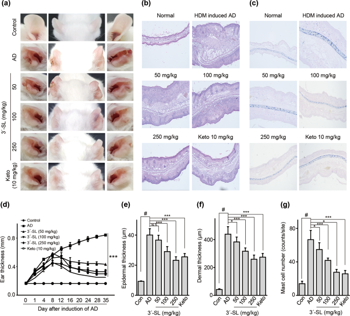
HDM-specific IgE28. To determine the effect of


3′-SL on AD progression, experimental AD was induced via treatment of mouse ears with HDM at 2-day intervals (Fig. 1, Supplementary Fig. 1b,c), or by treatment with 1%


2,4,-dinitrochlorobenzene (DNCB) at 7-day intervals (Fig. 2, Supplementary Fig. 1d,e). Furthermore, we checked whether 3′-SL prevents allergic sensitization, elicitation, or both. Firstly,


3′-SL was orally administered after elicitation phase (Fig. 1, Supplementary Fig. 1b) or sensitization phase (Supplementary Fig. 1d, 2) in HDM-induced AD mice model. Although mouse-ear


thickness increased upon treatment with HDM, 3′-SL significantly decreased the ear thickness in both elicitation phase (Figs. 1a, 2d) and sensitization (Supplementary Fig. 2a,d).


Furthermore, after elicitation (Fig. 1) or sensitization stage, macroscopic analysis revealed that HDM induced AD lesions, including erythema, oedema, scaling, and bleeding, which were


suppressed upon administration of 3′-SL (Fig. 1a, Supplementary Fig. 2d). Histological changes were analysed by haematoxylin-eosin, and Toluidine Blue staining, which indicated that


epidermal and dermal thickness increased upon HDM treatment, and intradermal mast cell infiltration significantly increased in comparison with the control. However, these HMD-mediated


changes were markedly decreased by 3′-SL oral administration (Fig. 1c,e–g Supplementary Fig. 2c,e–g). 1% DNCB induced AD also causes allergic contact dermatitis with both sensitization and


elicitation phases. As shown in Fig. 2 and Supplementary Fig. 3, 1% DNCB-induced AD phenotypes were also decreased by 3′-SL in sensitization and elicitation phases. Together, these results


suggested that 3′-SL suppresses HDM and 1% DNCB-induced AD lesions by decreasing epidermal and dermal thickness and mast cell infiltration in both sensitization and elicitation phase. 3′-SL


INHIBITS THE PRODUCTION OF IGE AND PRO-INFLAMMATORY CYTOKINES IN AD-INDUCED MOUSE SERUM Human AD patients shown high level of IgE and expressed various pro inflammatory cytokines28.


Therefore, To determine the mechanism underlying 3′-SL-mediated inhibition of AD progression, we used ELISA to measure plasma levels of IgE and pro-inflammatory cytokines in HDM and 1% DNCB


induced AD mice with 3′-SL oral administration. Serum IgE levels in the mice decreased upon 3′-SL administration (Fig. 3b,d) Furthermore, Plasma IL-1β, IL-6, and TNF-α levels were decreased


in 3′-SL-treated mice with AD (Fig. 3a,c). These results suggest that 3′-SL suppresses AD by reducing the production of IgE level and pro-inflammatory cytokines. 3′-SL BLOCKS HDM AND 1%


DNCB-INDUCED AD PATHOGENIC CYTOKINES VIA NF-ΚB INACTIVATION IN THE EAR To investigate the mechanism underlying 3′-SL-mediated suppression of HDM and 1% DNCB-induced AD, we determined the


transcript levels of AD-related inflammatory cytokines in the treated mouse ear tissue by qRT-PCR analysis (Fig. 4 and Supplementary Fig. 4). Th1 type cytokines (IFN-γ, TNF-α) and Th2 type


cytokines (IL-4, IL-5, IL-13) were upregulated in HDM- and 1% DNCB-induced mice ear tissue. However, oral administration of 3′-SL (100 and 250 mg/kg) inhibited expression of Th1 cytokines


(Fig. 4a and Supplementary Fig. 4a) IFN-gamma and (Fig. 4b and Supplementary Fig. 4b) TNF-α; and Th2 cytokines (Fig. 4c and Supplementary Fig. 4c) IL-4 (Fig. 4d and Supplementary Fig. 4d)


IL-5 and (Fig. 4e and Supplementary Fig. 4e) IL-13; and other AD-related cytokines (Fig. 4f and Supplementary Fig. 4f) IL-17 and (Fig. 4g and Supplementary Fig. 4g) Tslp, were also


downregulated by oral administration of 3′-SL in the AD induced mice. These results indicate that 3′-SL downregulates the production of cytokines produced by a T-cell subset in the ear


tissue of AD mice. NF-κB is a transcriptional regulator involved in various immune responses, including skin inflammatory diseases29. Many studies have reported that AD-related


pro-inflammatory cytokines are regulated by NF-κB29,30. Therefore, we investigated whether 3′-SL blocks the intranuclear translocation of p65 via immunofluorescence staining. As shown in


Supplementary Fig. 5, nuclear localization of p65 in the ear tissue of AD mice was suppressed via oral administration of 3′-SL in a dose dependent manner. These results indicate that 3′-SL


suppressed AD-related pro-inflammatory cytokines by blocking intranuclear p65 translocation and inhibiting NF-κB activation. 3′-SL ALLEVIATES THE SYSTEMIC IMMUNE RESPONSE BY INHIBITING T


CELL ACTIVATION Various studies have reported that AD progresses to systemic diseases, including autoimmune disorder, inflammatory bowel disease, and metabolic disease31,32. Therefore, we


investigated the expression of pro-inflammatory cytokine expression and production in Jurkat T cells. We initially investigated whether 3′-SL is cytotoxic to Jurkat T cells. As shown in Fig.


 5a, treatment of Jurkat T cells with 3′-SL at various concentrations for 24 h did not result in cytotoxic effects. Thus, an _in vitro_ analysis was performed wherein Jurkat T cells were


treated with 0 to 250 μM 3′-SL, in the presence of stimulant PMA/A23187 for 24 h, and then the transcript levels and production of pro-inflammatory cytokines IL-1β, IL-6, and TNF-α were


assessed via qRT-PCR and ELISA. As shown in Fig. 5b,c, upregulation and production of IL-1β, IL-6, and TNF-α by PMA/A23187 were drastically decreased by 3′-SL in a dose-dependent manner.


These results suggest that 3′-SL suppresses pro-inflammatory cytokines by inhibiting T cell activation. 3′-SL INDUCED REGULATORY T CELL DIFFERENTIATION Regulatory T cells suppress excessive


inflammatory responses and induce immunologic tolerance to self-antigens, thus inhibiting mast cell activation and IgE production33. Consistent with the results mentioned above, 3′-SL


suppressed mast cell infiltration and IgE production, and reduced T cell activation through blockade of pro-inflammatory cytokine secretion in Jurkat T cells. Therefore, we next determined


whether 3′-SL affects the frequency of Treg cells for both elicitation phase (Fig. 6a,b) and sensitization phase (Fig. 6c,d). Total cell number in the draining lymph nodes increased (Fig. 


6b,d; left panel) and the total number of CD4+ FoxP3+ cells decreased (Fig. 6b,d; right panel) in the HDM and 1% DNCB (Supplementary Fig. 6)-induced AD group in comparison with the control


group. However, the frequency (Fig. 6a,b; middle panel, Supplementary Fig. 6a,b; middle panel) and total number of CD4 + Foxp3+ cells (Fig. 6b, Supplementary Fig. 6b; right panel) in the


draining lymph node was influenced by oral 3′-SL administration. These results suggest that 3′-SL induced the Treg population in the draining lymph node. To verify the effect of 3′-SL on


Treg differentiation, we investigated whether 3′-SL treatment enhances Treg differentiation _in vitro_. CD4 + T cells were isolated from the spleen of C57BL/6 mice and cultured in the


presence and absence of TGF-β or co-treated with 3′-SL for 3 days. As shown in Fig. 6e,f, co-treatment with 3′-SL and TGF-β significantly increased the Treg population in comparison with the


control group and TGF-β-treated group. These results indicate that 3′-SL attenuates progression of AD by promoting differentiation of Tregs. 3′-SL ADMINISTRATION INCREASES INCIDENCE OF


_BIFIDOBACTERIUM_ 3′-SL is known to promote the outgrowth of _Bifidobacterium_. Moreover, _Bifidobacterium_ is a well-known probiotics for AD34,35. To check whether oral administration of


3′-SL modulates _Bifidobacterium_ in normal conditions, we first determined levels of _Bifidobacterium_ after administration of 3′-SL oral for 35 day in WT mouse. _Bifidobacterium_,


especially_, Bifidobacterum bifidium_, were gradually increased by 3′-SL oral administration in dose dependent manner (Fig. 7a). Subsequently, we checked effect of 3′-SL modulate on


_Bifidobacterium_ in the AD mouse models. Although _Bifidobacterium_ were decreased in HDM (Figs. 7b) and 1% DNCB (Fig. 7c) induced AD pathogenesis after elicitation phase,


_Bifidobacterium_, especially_, Bifidobacterum bifidium_, were increased by 3′-SL oral administration with rescuing AD phenotypes. These results indicate that 3′-SL can function as a


prebiotic to modulate _Bifidobacterium_ levels. DISCUSSION Prebiotics are ingredients that have beneficial effects on the host by selectively stimulating intestinal bacteria that can improve


host health. Gradually increasing evidence has indicated the beneficial effect of prebiotics on the treatment of patients with AD, which is a chronic inflammatory disease characterized by


sensitivity to various allergens and several genetic, environmental, and immunological factors4. The common immunological characteristics of patients with AD are mast cell infiltration at


the site of the lesion and elevated serum IgE levels8,36 Furthermore, Th2 and Th17 immune responses resulting from immune imbalance lead to the secretion of Th2 and Th17 cytokines (e.g.,


IL-4, IL-5, IL-13, IL17)5,6,37. This study shows that 3′-SL attenuates AD pathogenesis by inducing Tregs, thus inhibiting the production of the T cell subset (Th1, Th2, Th17) cytokines


(e.g., IL-4, IL-5, IL-13, IL-17, IFN-γ, TNF-α, and Tslp) and reducing T cell activation. Furthermore, 3′-SL reduced the production of IgE in mouse serum and mast cell infiltration in


sensitization and elicitation phases to the ear. Human breast milk contains various bioactive molecules with developmental and protective functions. 3′-SL is one of various human milk


oligosaccharides and prebiotics that comprise monosaccharide N-acetylneuraminic acid linked to the galactosyl subunit of lactose at the 3′ position. Particularly, 3′-SL exerts


anti-inflammatory effects and supports immune homeostasis23,24. Furthermore, 3′-SL promotes the growth and activity of beneficial intestinal bacteria and protects against arthritis. However,


no studies have focused on the effect of 3′-SL on skin inflammation. In AD, T lymphocytes are activated and predominantly infiltrate the site where the AD lesions are located38. Th1 cells


produce IFN-γ and TNF for macrophage activation, resulting in a delayed immune response. In contrast, Th2 cells produce IL-4, IL-5, and IL-13 to increase IgE synthesis and promote mast


cell-induced type 1 hypersensitivity5,30,37. IL-1β is involved in the progression of AD and is a prominent inflammatory cytokine secreted at high concentrations by immune cells and


epithelial cells in lesions and the blood of patients with AD. Herein, oral administration of 3′-SL blocked the secretion of IL-1β, IL-6, and TNF-α. Furthermore, the Th1 (IFN-γ, TNF-α) and


Th2 (IL-4, IL-5, IL-13) type cytokines in AD lesions in mice were markedly downregulated upon oral 3′-SL administration. Recent evidence suggests that IL-1β regulates the production of Tslp,


resulting in AD progression39. Tslp is also known as the “master switch” of allergic inflammatory reactions because it affects mast and T cells, leading to skin inflammation, and basophils,


and eosinophils leading to both innate and acquired immune responses and the induction of Th2 immune responses40. Tslp is an important factor in the pathogenesis of AD39,41. In patients


with AD, Tslp production is increased in the skin and induces Th2 inflammation40. Herein, Tslp was markedly downregulated upon treatment with 3′-SL in mouse ear tissue. Secretion of Th cell


subset cytokines by T cell activation is modulated by transcription factors, including NF-κB, which regulates genes associated with various inflammatory responses29,30,42. NF-κB plays an


important role in innate/adaptive immune responses and chronic inflammatory responses, particularly in Th1 responses, and regulates inflammation resulting from Th2 cell differentiation and


activation30. NF-κB is a heterodimeric form of p50/p65, which is present in the cytoplasm and regulates various inflammatory genes upon intranuclear translocation of p65 in various


inflammatory conditions42. Herein, 3′-SL downregulated proinflammatory cytokines via suppressed T cell activation in Jurkat T cells. In particular, the mechanism underlying the effects of


3′-SL is associated with NF-κB transcriptional regulation via the blockade of intranuclear p65 translocation. Activated T cells are suppressed by Tregs, which, in turn, are regulated by


NF-κB. Tregs have recently been shown to play a role in immune disorders, including AD pathogenesis18,43. Tregs secrete immunosuppressive cytokines including IL-10 and TGF-β to suppress


excessive inflammatory responses, resulting in blocked secretion of cytokines including IL-1β, IL-6, IL-4, IL-17, TNF-α, and Tslp13,16,18. Furthermore, Tregs induce immunological tolerance


to self-antigens. Tregs inhibit mast cell and T cell activation during allergic reactions and reduce IgE production by promoting isotype switching of B cells11,33. However, high levels of


IgE inhibit IL-10 signaling and Treg differentiation in patients with allergy44. Many studies have indicated the importance of promoting the differentiation, proliferation, and activity of


Treg in allergy patients11,15. 3′-SL acts as a prebiotic and leads to changes in the intestinal microbiota profiling. Some studies indicate that oral administration of 3′-SL increases the


microbiota, including _Bifidobacterium_34. Especially, oral administration of these microbiota attenuated AD progression35. Interestingly, oral administration of _Bifidobacterium_ induces


the Treg population with polysaccharide A or outer membrane vesicles form _Bacteriodes fragilis_45. In our study, we checked whether 3′-SL increased _Bifidobacterium_ in AD pathogenesis.


Although general _Bifidobacterium_ were decreased in DNCB and HDM-induced AD pathogenesis, the oral administration of 3′-SL increased most _Bifidobacterium_, especially _Bifidobacterium


bifidum_, along with reduced AD pathogenesis. Herein, the total cell number and frequency in the draining lymph node remained unchanged in the AD group; however, the total number of Tregs


was lesser than that in the control group. Moreover, the frequency and total number of Tregs in the draining lymph node was increased upon oral 3′-SL administration. Treg induction


reportedly attenuates autoimmune diseases, including AD and T1D19,46. Furthermore, _ex vivo_ analysis of T cell differentiation revealed that treatment with 3′-SL enhanced the CD4+ Foxp3 + T


cell population. In conclusion, this study shows that oral 3′-SL administration reduces the progression of HDM and 1% DNCB-induced AD by reducing T cell activation, enhancing T cell


differentiation into Tregs, and increasing the _Bifidobacterium_ population. Furthermore, 3′-SL regulates the expression of cytokines produced by Th1 cells (IFN-γ, TNF-α), Th2 cells (IL-4,


IL-5, IL-13) and Th17 cells (IL-17) by blocking the intranuclear translocation of p65 in the ear tissue of HDM and 1% DNCB induced AD model mice. Moreover, The doses of 50, 100, and 250 


mg/kg in mouse is similar to 17.5, 35.1, and 87.5 mg/kg once every two days in human. If consumed every day, then the sufficient dose would be 8.75, 17.5 and 43.75 mg/kg per day.54 The


present results suggest that 3′-SL is a potential therapeutic agent for AD and its mechanism involves regulation of Treg differentiation, inflammation, and _Bifidobacterium_ population.


MATERIALS AND METHODS ANIMALS Eight-week-old male BALB/c and C57BL/6 mice weighing 18–20 g were purchased from DBL, Chungbuk, Korea and housed under standard conditions (temperature, 22–25 


°C; 12 h photoperiod). All animal experiments were approved by the Animal Care and Use Committee of the University of Ajou and Global campus, Kyung Hee University (Protocol number:


KHGASP-17–040). All _in vivo_ experiments were performed according to the guidelines of the National Institutes of Health and in accordance with relevant guidelines and regulations.


ESTABLISHMENT OF AN AD MOUSE MODEL WITH HDM AND 1% DNCB In the HDM (_Dermatophagoides farinae_ extract, Greer Laboratories, Lenoir, NC, USA)-induced atopic dermatitis model, both surfaces of


the ear lobes were very gently stripped three times with Tegaderm (Supplementary Fig. 1c). After stripping, 20 μL of HDM (10 mg/mL) was painted on each ear. The application of HDM was


repeated 3 times per week for 5 weeks. Experimental AD was induced in 8-week-old male BALB/c mice through serial exposure to _D. farinae_ extract (DFE) and DNCB around the ear (Supplementary


Fig. 1b), as previously described47. 3′-Sialyllactose was purchased from SYNOVIZEN Inc. (Seoul, South Korea). Mice were randomly separated into six groups: control, AD-induction,


AD-induction + 50, 100, and 250 mg/kg of 3′-SL and Positive control (Ketotifen 10 mg/kg). Earlobe surfaces were stripped using surgical tape (Nichiban, Tokyo, Japan), and 1% DNCB was applied


to each earlobe; DEF (10 mg/mL) was also applied after 4 d on the same site. DEF/DNCB was administered once a week for 5 weeks. 3′-SL (50, 100 and 250 mg/kg) was orally administered for 5


weeks during AD induction. Ear thickness was measured using a dial thickness gauge (Kori Seiki MFG, Co., Tokyo, Japan) after 24 h of DEF/DNCB administration and every 4 days after induction


of AD. Blood samples were collected from mice after 35 d and stored at –80 °C until further use. CELL CYTOTOXICITY ANALYSIS The cytotoxicity of 3′-SL on Jurkat T cells, cultured in RPMI


medium containing 10% foetal bovine serum (FBS), 50 U/mL penicillin, and 50 µg/mL streptomycin, was assessed using the EZ-CyTox Cell viability assay kit (Dogen, Seoul, South Korea) following


the manufacturer’s protocol. Briefly, cells were seeded into 96-well culture plate at 1 × 10⁴ cells/well. 3′-SL was supplemented in the cell culture media at 10, 50, 100, and 250 μM, and


cells were cultured for 24 h. Thereafter, the WST-1 solution (2-(4-iodophenyl)-3-(4-nitrophenyl)-5-(2,4 disulfophenyl)-2H-tetrazolium) was mixed with serum-free RPMI (1:10 v/v) and added to


each well for 2 h in the dark. Cytotoxicity was determined at 450 nm using a microplate reader VICTOR X3 (PerkinElmer, Waltham, MA, USA). QUANTITATIVE REVERSE TRANSCRIPTASE PCR (QRT-PCR)


ANALYSIS Total RNA was isolated from mouse ear tissue and Jurkat T cells using TRIzol reagent (Molecular Research Center Inc., Cincinnati, OH, USA) in accordance with the manufacturer’s


instructions. cDNA was synthesized using 1 µg of total mRNA and amplified with target gene primers (Table S1) via PCR (Intron Biotechnology, Gyeonggi-do, South Korea). Relative mRNA


expression levels were determined using the SYBR premix Ex Taq (TaKaRa Bio, Shiga, Japan) and normalized to those of _GAPDH_. To detect _Bifidobacterium_, fresh faecal samples were collected


from mice and immediately stored at −150 °C until processing. Faecal DNA was isolated using the FastDNA Spin kit (MP Biomedicals, Santa Ana, CA, USA). PCR amplification was performed with


target gene primer (Table S2)48,49 via qRT-PCR. Quantification values were calculated by the 2−ΔΔCt method relative to total bacteria 16 S rDNA amplification. ENZYME-LINKED IMMUNOSORBENT


ASSAY (ELISA) IL-1β, IL-6, TNF-α, and IgE production was quantified using an ELISA kit (Koma Biotech, Seoul, South Korea) in accordance with the manufacturer’s instructions. Absorbance for


each cytokine and IgE was determined at 450 nm using a microplate reader (VICTOR X3; PerkinElmer, Waltham, MA, USA). IMMUNOFLUORESCENCE ASSAYS Mouse-ear tissue was cut into 5-µm-thick


sections, which were incubated with rabbit anti-p65 antibody (1:200) (8242; Cell Signaling Technology) overnight at 4 °C. After washing with PBS, the sections were incubated with goat


anti-rabbit secondary antibody (ab150081; Abcam, Cambridge, UK) conjugated with Alexa Fluor 488 for 2 h at room temperature. Thereafter, slides were washed with PBS and incubated with DAPI


solution (4,6-diamidino-2-phenylindole) (DAPI; Invitrogen, San Francisco, CA, USA) for 10 min at room temperature. Mouse ear tissue was imaged using Axioscan (Zeiss) at the Three-dimensional


immune system core facility of Ajou University. ANTIBODY STAINING AND FLOW CYTOMETRIC ANALYSES Draining lymph node samples were obtained from BALB/c mice. Single cells were isolated via


cervical dislocation, washed with PBS containing 10% FBS, and surface stained for CD4 using FITC-conjugated anti-CD4 antibody (300506; BioLegend, San Diego, CA, USA). Subsequently, cells


were resuspended in fixation/permeabilization buffer for 12 h and stained for intracellular Foxp3 using anti-Foxp3 APC antibody (17-5773-82; Thermo Fisher Scientific, Waltham, MA, USA)


following the manufacturer’s instructions. Each sample was analysed using a FACS MACSQuant VYB flow cytometer (Bergisch Gladbach, Germany). T CELL PROLIFERATION ASSAY Spleen samples were


obtained from C57BL/6 mice. Single cells were isolated via cervical dislocation; CD4+ T cells were negatively selected using Anti-Biotin MicroBeads (130-090-485; Miltenyi). CD4+ T cells were


re-suspended (106 cells/mL) in complete culture media comprising TexMacs medium supplemented with 10% FBS, 4 mM L-glutamine, 50 U/mL penicillin, and 50 μg/mL streptomycin; the cells were


then incubated in 24-well plates pre-coated with 5 μg/mL anti-mouse CD3 monoclonal antibody (100201; Biolegend) and 1 μg/mL anti-mouse CD28 antibody (122022; Biolegend) and treated with


3′-SL (50, 100, 250 μM) and TGF-β (10 ng) for 3, 6, and 9 d. The Treg population was assessed via FACS. STATISTICAL ANALYSIS Data are presented as mean ± SD values. Differences between the


control and treatment groups were evaluated using one-way ANOVA with Dunnett’s post-hoc multiple comparison tests. Statistical significance was determined at _P_ < 0.05. Statistical


analysis was performed using GraphPad Prism 5 software (GraphPad, San Diego, CA, USA)49. REFERENCES * Markowiak, P. & Śliżewska, K. Effect of Probiotics, Prebiotics, and Synbiotics on


Human Health. _Nutrients._ 9, 9 (2017). Article  CAS  Google Scholar  * Carlson, J. L., Erickson, J. M., Lloyd, B. B. & Slavin, J. L. Health Effects and Sources of Prebiotic Dietary


Fiber. _Curr. Dev. Nutr._ 2, 3 (2018). Article  CAS  Google Scholar  * Kiousi, D. E. _et al_. Probiotics in Extraintestinal Diseases: Current Trends and New Direction. _Nutrients._ 11, 4


(2019). Article  CAS  Google Scholar  * Lyons, J. J., Milner, J. D. & Stone, K. D. Atopic dermatitis in children: clinical features, pathophysiology, and treatment. Immunol. _Allergy.


Clin. North. Am_ 35, 161–83 (2015). Article  Google Scholar  * Brandt, E. B. & Sivaprasad, U. T. 2 Cytokines and Atopic Dermatitis. _J. Clin. Cell. Immunol_ 2, 110 (2010). Google Scholar


  * Klonowska, J., Gleń, J., Nowicki, R. J. & Trzeciak, M. New Cytokines in the Pathogenesis of Atopic Dermatitis—New Therapeutic Targets. _Int. J. Mol. Sci_ 19, 3086 (2018). Article 


PubMed Central  CAS  Google Scholar  * Gandhi, N. A., Pirozzi, G. & Graham, N. M. H. Commonality of the IL-4/IL-13 pathway in atopic diseases. _Expert. Rev. Clin. Immunol_ 13, 425–437


(2017). Article  CAS  PubMed  Google Scholar  * Galli, S. J. & Tsai, M. IgE and mast cells in allergic disease. _Nat. Med_ 18, 693–704 (2012). Article  CAS  PubMed  PubMed Central 


Google Scholar  * Zhang, B. _et al_. Human mast cell degranulation and preformed TNF secretion require mitochondrial translocation to exocytosis sites: relevance to atopic dermatitis. _J.


Allergy. Clin. Immunol_ 127, 1522–31 (2011). Article  CAS  PubMed  PubMed Central  Google Scholar  * Kawakami, T. _et al_. Mast cells in atopic dermatitis. _Curr. Opin. Immunol_ 21, 666–678


(2019). Article  CAS  Google Scholar  * Sakaguchi, S., Yamaguchi, T., Nomura, T. & Ono, M. Regulatory T cells and immune tolerance. _Cell_ 133, 775–87 (2008). Article  CAS  PubMed 


Google Scholar  * Nie, J. _et al_. FOXP3(+) Treg Cells and Gender Bias in Autoimmune Diseases. _Front. Immunol_ 6, 493 (2015). Article  PubMed  PubMed Central  CAS  Google Scholar  *


Fyhrquist, N. _et al_. Foxp3+ cells control Th2 responses in a murine model of atopic dermatitis. _J. Invest. Dermatol_ 132, 1672–80 (2012). Article  CAS  PubMed  Google Scholar  * Nadeau,


K. _et al_. Ambient air pollution impairs regulatory T-cell function in asthma. _J. Allergy. Clin. Immunol_ 126, 845–852 (2010). Article  CAS  PubMed  Google Scholar  * Rivas, M. N. &


Chatila, T. A. Regulatory T cells in Allergic Diseases. _J. Allergy. Clin. Immunol_ 138, 639–652 (2016). Article  PubMed Central  CAS  Google Scholar  * Roesner, L. M. _et al_. Foxp3(+)


regulatory T cells are expanded in severe atopic dermatitis patients. _Allergy_ 70, 1656–60 (2015). Article  CAS  PubMed  Google Scholar  * Kupper, T. S. & Fuhlbrigge, R. C. Immune


surveillance in the skin: mechanisms and clinical consequences. _Nat. Rev. Immunol_ 4, 211–22 (2014). Article  CAS  Google Scholar  * Agrawal, R., Wisniewski, J. & Woodfolk, J. A. The


Role of Regulatory T cells in atopic dermatitis. _Curr. Probl. Dermatol_ 41, 112–124 (2011). Article  CAS  PubMed  PubMed Central  Google Scholar  * Ogino, H. _et al_. Regulatory T cells


expanded by rapamycin _in vitro_ suppress colitis in an experimental mouse model. _J. Gastroenterol_ 47, 366–76 (2012). Article  ADS  CAS  PubMed  Google Scholar  * Galgani, M., Rosa, V. D.,


Cava, A. L. & Matarese, G. Role of Metabolism in the Immunobiology of Regulatory T Cells. _J. Immunol_ 197, 2567–2575 (2016). Article  CAS  PubMed  Google Scholar  * Nasri, H. &


Rafieian-Kopaei, M. Metformin: Current knowledge. _J. Res. Med. Sci_ 19, 658–64 (2014). PubMed  PubMed Central  Google Scholar  * Fischer, K. E. _et al_. Health Effects of Long-Term


Rapamycin Treatment: The Impact on Mouse Health of Enteric Rapamycin Treatment from Four Months of Age throughout Life. _PLoS. One_ 10, e0126644 (2015). Article  PubMed  PubMed Central  CAS


  Google Scholar  * Luo, J. _et al_. An integrated membrane system for the biocatalytic production of 3′-sialyllactose from dairy by-products. _Bioresour. Technol_ 166, 9–16 (2014). Article


  CAS  PubMed  Google Scholar  * Donovan, S. M. & Comstock, S. S. Human Milk Oligosaccharides Influence Neonatal Mucosal and Systemic Immunity. _Ann. Nutr. Metab_ 69, 42–51 (2016).


Article  PubMed  Google Scholar  * Perdijk, O. _et al_. Sialyllactose and Galactooligosaccharides Promote Epithelial Barrier Functioning and Distinctly Modulate Microbiota Composition and


Short Chain Fatty Acid Production. _In Vitro. Front. Immunol._ 10, 94 (2019). Article  CAS  PubMed  Google Scholar  * Jeon, J. _et al_. 3′-Sialyllactose protects against osteoarthritic


development by facilitating cartilage homeostasis. _J. Cell. Mol. Med_ 22, 57–66 (2018). Article  CAS  PubMed  Google Scholar  * Kang, L. J. _et al_. 3′-Sialyllactose as an inhibitor of p65


phosphorylation ameliorates the progression of experimental rheumatoid arthritis. _Br. J. Pharmacol_ 175, 4295–4309 (2018). Article  CAS  PubMed  PubMed Central  Google Scholar  * Martel,


B., Lovato, P., Bäumer, W. & Olivry, T. Translational Animal Models of Atopic Dermatitis for Preclinical Studies. _Yale. J. Biol. Med_ 90(3), 389–402 (2017). CAS  PubMed  PubMed Central


  Google Scholar  * Wullaert, A., Bonnet, M. C. & Pasparakis, M. NF-κB in the regulation of epithelial homeostasis and inflammation. _Cell. Res_ 21, 146–158 (2011). Article  CAS  PubMed


  Google Scholar  * Tenda, Y. _et al_. Hyperresponsive TH2 cells with enhanced nuclear factor-kappa B activation induce atopic dermatitis-like skin lesions in Nishiki-nezumi Cinnamon/Nagoya


mice. _J. Allergy. Clin. Immunol_ 118, 725–33 (2016). Article  CAS  Google Scholar  * Thijs, J. L. _et al_. Serum biomarker profiles suggest that atopic dermatitis is a systemic disease. _J.


Allergy. Clin. Immunol_ 141, 1523–1526 (2018). Article  PubMed  Google Scholar  * Darlenski, R., Kazandjieva, J., Hristakieva, E. & Fluhr, J. W. Atopic dermatisis as a systemic disease.


_Clin. Dermatol_ 32, 409–13 (2014). Article  PubMed  Google Scholar  * Gri, G. _et al_. CD4+ CD25+ regulatory T cells suppress mast cell degranulation and allergic responses through


OX40-OX40L interaction. _Immunity_ 29, 771–8 (2008). Article  CAS  PubMed  PubMed Central  Google Scholar  * Perdijk, O. _et al_. Sialyllactose and Galactooligosaccharides Promote Epithelial


Barrier Functioning and Distinctly Modulated Microbiota Composition and Short Chain Fatty Acid Production _in Vitro_. _Front. immunol_ 10, 94 (2019). Article  CAS  PubMed  PubMed Central 


Google Scholar  * Wollina, U. Microbiome in atopic dermatitis. _Clin. Cosmet. Investig. Dermatol_ 10, 51–56 (2017). Article  CAS  PubMed  PubMed Central  Google Scholar  * Liu, F. T.,


Goodarzi, H. & Chen, H. Y. IgE, mast cells, and eosinophils in atopic dermatitis. _Clin. Rev. Allergy. Immunol_ 41, 298–310 (2011). Article  CAS  PubMed  Google Scholar  * Di, Z. H. _et


al_. T Helper 1 and T Helper 2 Cytokines Differentially Modulate Expression of Filaggrin and its Processing Proteases in Human Keratinocytes. _Chin. Med. J (Engl)_ 129, 295–303 (2016).


Article  Google Scholar  * Werfel, T. _et al_. Cellular and molecular immunologic mechanisms in patients with atopic dermatitis. _J. Allergy. Clin. Immunol_ 138, 336–49 (2016). Article  CAS


  PubMed  Google Scholar  * Bernard, M. _et al_. IL-1β induces thymic stromal lymphopoietin and an atopic dermatitis-like phenotype in reconstructed healthy human epidermis. _J. Pathol_ 242,


234–245 (2017). Article  CAS  PubMed  Google Scholar  * Liu, Y. J. Thymic stromal lymphopoietin: master switch for allergic inflammation. _J. Exp. Med_ 203, 269–273 (2006). Article  PubMed


  PubMed Central  Google Scholar  * Cianferoni, A. & Spergel, J. The importance of TSLP in allergic disease and its role as a potential therapeutic target. _Expert. Rev. Clin. Immunol_


10, 1463–1474 (2014). Article  CAS  PubMed  PubMed Central  Google Scholar  * Tanaka, A. _et al_. Topical application with a new NF-kappaB inhibitor improves atopic dermatitis in NC/NgaTnd


mice. _J. Invest. Dermatol_ 127, 855–63 (2017). Article  CAS  Google Scholar  * Grinberg-Bleyer, Y. _et al_. Alternative NF-κB Pathway in Regulatory T Cell Homeostasis and Suppressive


Function. _J. Immunol_ 200, 2362–2371 (2018). Article  CAS  PubMed  Google Scholar  * Saito, M. _et al_. Defective IL-10 signaling in hyper-IgE syndrome results in impaired generation of


tolerogenic dendritic cells and induced regulatory T cells. _J. Exp. Med_ 208, 235–249 (2011). Article  ADS  CAS  PubMed  PubMed Central  Google Scholar  * Round, J. L. & Mazmanian, S.


K. Inducible Foxp3+regulatory T-cell development by a commensal bacterium of the intestinal microbiota. _Proc. Natl. Acad. Sci. USA_ 107, 12204–9 (2010). Article  ADS  CAS  PubMed  PubMed


Central  Google Scholar  * Spence, A., Klementowicz, J. E., Bluestone, J. A. & Tang, Q. TargetingTreg signaling for the treatment of autoimmune diseases. _Curr. Opin. Immunol_ 37, 11–20


(2015). Article  CAS  PubMed  PubMed Central  Google Scholar  * Choi, E. J. _et al_. Heat-Killed Enterococcus faecalis EF-2001 Ameliorates Atopic Dermatitis in a Murine Model. _Nutrients_ 8,


146 (2016). Article  PubMed  PubMed Central  CAS  Google Scholar  * Kwon, M. S. _et al_. _Lactobacillus sakei_ WIKIM30 Ameliorates Atoipic Dermatitis-Like Skin Lesions by Inducing


Regulatory T cells and Alteraing Gut Microbiota Structure in Mice. _Front Immunol_ 9, 1–11 (2018). Article  CAS  Google Scholar  * Bergstrom, A. _et al_. Introducing GUt Low-Density Array


(GULDA)-a validated approach for qPCR-based intestinal microbial community analysis. _FEMS Microbiol Lett_ 337, 38–47 (2012). Article  CAS  PubMed  Google Scholar  Download references


ACKNOWLEDGEMENTS This work was supported by the Korea Health Technology R&D Project through the Korea Health Industry Development Institute (HI16C0992). This work was supported by the


National Research Foundation of Korea (NRF) grant funded by the Korea government (2019R1A6A1A03032869, 2018R1C1B3001650, 2018R1C1B6007038 and SRC2017R1A5A1014560). This work was carried out


with the support of “Cooperative Research Program for Agriculture Science and Technology Development (Project No. PJ01330503)” Rural Development Administration, Republic of Korea. This work


was funded by a Korea Institute of Science and Technology intramural grant (2Z06220 and 2Z06130). AUTHOR INFORMATION Author notes * These authors contributed equally: Li-Jung Kang and


Eunjeong Oh. AUTHORS AND AFFILIATIONS * Department of Biomedical Sciences, Ajou University Graduate School of Medicine, Suwon, 16499, Republic of Korea Li-Jung Kang, Eunjeong Oh, Chanmi Cho,


 Jimin Jeon, Hyemi Lee, Sangil Choi, Seong Jae Han, Jiho Nam & Siyoung Yang * Department of Pharmacology, Ajou University School of Medicine, Suwon, 16499, Republic of Korea Li-Jung


Kang, Eunjeong Oh, Chanmi Cho, Jimin Jeon, Hyemi Lee, Sangil Choi, Seong Jae Han, Jiho Nam & Siyoung Yang * CIRNO, Sungkyunkwan University, Suwon, 16419, Republic of Korea Li-Jung Kang, 


Eunjeong Oh, Chanmi Cho, Jimin Jeon, Hyemi Lee, Sangil Choi, Seong Jae Han, Jiho Nam, Hye Young Kim & Siyoung Yang * Department of Microbiology and Immunology, Yonsei University College


of Medicine, Seoul, 03722, Korea HoKeun Kwon * Korea Institute of Science & Technology (KIST) Gangneung Institute of Natural Products, Gangwon-do, 25451, Republic of Korea Choong-Gu Lee


* Department of Life Science, Chung-Ang University, Seoul, 06974, Republic of Korea Chi-une Song & Seong-il Eyun * Synovizen Inc, Seoul, 06621, Republic of Korea Hyunho Jung * Laboratory


of mucosal immunology, Department of Biomedical Science, Seoul National University College of Medicine, Seoul, 03080, Republic of Korea Hye Young Kim * East-West Medical Research Institute,


Medical Science Research Institute, Kyung Hee University, Seoul, 02447, Republic of Korea Eun-Jung Park * Department of Physical Education, College of Education, Daegu Catholic University,


Gyeongsan, 38430, Republic of Korea Eun-Ju Choi * Department of Anatomy, School of Medicine, Kyungpook National University, Daegu, Republic of Korea Jooyoung Kim Authors * Li-Jung Kang View


author publications You can also search for this author inPubMed Google Scholar * Eunjeong Oh View author publications You can also search for this author inPubMed Google Scholar * Chanmi


Cho View author publications You can also search for this author inPubMed Google Scholar * HoKeun Kwon View author publications You can also search for this author inPubMed Google Scholar *


Choong-Gu Lee View author publications You can also search for this author inPubMed Google Scholar * Jimin Jeon View author publications You can also search for this author inPubMed Google


Scholar * Hyemi Lee View author publications You can also search for this author inPubMed Google Scholar * Sangil Choi View author publications You can also search for this author inPubMed 


Google Scholar * Seong Jae Han View author publications You can also search for this author inPubMed Google Scholar * Jiho Nam View author publications You can also search for this author


inPubMed Google Scholar * Chi-une Song View author publications You can also search for this author inPubMed Google Scholar * Hyunho Jung View author publications You can also search for


this author inPubMed Google Scholar * Hye Young Kim View author publications You can also search for this author inPubMed Google Scholar * Eun-Jung Park View author publications You can also


search for this author inPubMed Google Scholar * Eun-Ju Choi View author publications You can also search for this author inPubMed Google Scholar * Jooyoung Kim View author publications You


can also search for this author inPubMed Google Scholar * Seong-il Eyun View author publications You can also search for this author inPubMed Google Scholar * Siyoung Yang View author


publications You can also search for this author inPubMed Google Scholar CONTRIBUTIONS L.J.K., E.O., J.K., S.I.E. and S.Y. were in charge with conception, design, analysis, and


interpretation of data. L.J.K., C.C., J.J., H.L., S.C., S.J.H., E.O., J.N. and E.J.C. performed the _in vivo_ and _in vitro_ experiments. H.J., H.Y.K., E.J.P., C.U.S. and E.J.C. contributed


reagents, materials, and analytical tools for the study. H.K. and C.G.L. performed the microbiome analysis. L.J.K., J.K., S.I.E. and S.Y. wrote the paper and L.J.K. and S.Y. have full access


to overall data and takes responsibility for the integrity and accuracy of the data analysis. CORRESPONDING AUTHORS Correspondence to Jooyoung Kim, Seong-il Eyun or Siyoung Yang. ETHICS


DECLARATIONS COMPETING INTERESTS The authors declare no competing interests. ADDITIONAL INFORMATION PUBLISHER’S NOTE Springer Nature remains neutral with regard to jurisdictional claims in


published maps and institutional affiliations. SUPPLEMENTARY INFORMATION SUPPLYMENTARY INFORMATION. RIGHTS AND PERMISSIONS OPEN ACCESS This article is licensed under a Creative Commons


Attribution 4.0 International License, which permits use, sharing, adaptation, distribution and reproduction in any medium or format, as long as you give appropriate credit to the original


author(s) and the source, provide a link to the Creative Commons license, and indicate if changes were made. The images or other third party material in this article are included in the


article’s Creative Commons license, unless indicated otherwise in a credit line to the material. If material is not included in the article’s Creative Commons license and your intended use


is not permitted by statutory regulation or exceeds the permitted use, you will need to obtain permission directly from the copyright holder. To view a copy of this license, visit


http://creativecommons.org/licenses/by/4.0/. Reprints and permissions ABOUT THIS ARTICLE CITE THIS ARTICLE Kang, LJ., Oh, E., Cho, C. _et al._ 3′-Sialyllactose prebiotics prevents skin


inflammation via regulatory T cell differentiation in atopic dermatitis mouse models. _Sci Rep_ 10, 5603 (2020). https://doi.org/10.1038/s41598-020-62527-5 Download citation * Received: 11


December 2019 * Accepted: 12 March 2020 * Published: 27 March 2020 * DOI: https://doi.org/10.1038/s41598-020-62527-5 SHARE THIS ARTICLE Anyone you share the following link with will be able


to read this content: Get shareable link Sorry, a shareable link is not currently available for this article. Copy to clipboard Provided by the Springer Nature SharedIt content-sharing


initiative