Innovative ochre processing and tool use in China 40,000 years ago
- Select a language for the TTS:
- UK English Female
- UK English Male
- US English Female
- US English Male
- Australian Female
- Australian Male
- Language selected: (auto detect) - EN
Play all audios:
Homo sapiens was present in northern Asia by around 40,000 years ago, having replaced archaic populations across Eurasia after episodes of earlier population expansions and
interbreeding1,2,3,4. Cultural adaptations of the last Neanderthals, the Denisovans and the incoming populations of H. sapiens into Asia remain unknown1,5,6,7. Here we describe Xiamabei, a
well-preserved, approximately 40,000-year-old archaeological site in northern China, which includes the earliest known ochre-processing feature in east Asia, a distinctive miniaturized
lithic assemblage with bladelet-like tools bearing traces of hafting, and a bone tool. The cultural assembly of traits at Xiamabei is unique for Eastern Asia and does not correspond with
those found at other archaeological site assemblages inhabited by archaic populations or those generally associated with the expansion of H. sapiens, such as the Initial Upper
Palaeolithic8,9,10. The record of northern Asia supports a process of technological innovations and cultural diversification emerging in a period of hominin hybridization and
admixture2,3,6,11.
AMS 14C and OSL age data used in Fig. 2 are presented in Supplementary Tables C1, C3, respectively. The data from X-ray diffraction spectra, Raman spectra, high-temperature magnetic
susceptibility measurements, and magnetic component analysis of coercivity distributions used in Extended Data Fig. 6 can be downloaded at https://doi.org/10.5061/dryad.9s4mw6mj0. Other data
generated during this study are included in the Article, Extended Data and Supplementary Information, and/or are available from the corresponding author/s on reasonable request.
CQL code for the Bayesian age model is provided in Supplementary Table C4
We thank Y. Lefrais (IRAMAT-CRP2A, UMR 5060 CNRS) and F. Orange for assistance with SEM–EDS analyses; A. Queffelec (PACEA UMR 5199) and L. Geis (PACEA UMR 5199) for assistance with the EDXRF
analyses and the 3D imaging; C. X. Zhang, B. Hu, M. L. Zhou, J. H. Li, Y. Liu, S. H. Yang, X. G. Li, Y. Chen, J. Yuan, Z. S. Shen, S. Zhang and Z. X. Jiang for assistance with the sediment
analysis; B. Xu and Y. Li for discussions on dating results; and R. P. Tang and F. X. Huan for assistance with figure preparation. Financial support for this research was provided by the
National Natural Science Foundation of China (41888101, 42177424, 41977380, 42072212 and 41690112), the Strategic Priority Research Program of Chinese Academy of Sciences (XDB26000000), the
Key Research Program of the Institute of Geology and Geophysics, Chinese Academy of Sciences (IGGCAS-201905), the State Key Laboratory of Loess and Quaternary Geology, Institute of Earth
Environment (SKLLQGZR2002), the Youth Innovation Promotion Association of Chinese Academy of Sciences (2020074), the Humboldt Foundation, and the Max Planck Society. A.O. was supported by
the Spanish MICIU/Feder (PGC2018-093925-B-C32), the Catalan AGAUR (SGR2017-1040) and the Univ. Rovira i Virgili (2019-PFR-URV-91) in the context of a MICIN ‘María de Maeztu’ excellence
accreditation (CEX2019-000945). D.E.R. was funded by the Fyssen Foundation, France, and the Juan de la Cierva-Formación Research Fellowship (FJC2018-035605-I; Ministerio de Ciencia e
Innovación, Spain). F.d. was funded by the Research Council of Norway through its Centre of Excellence funding scheme (SFF Centre for Early Sapiens Behaviour–Sapien CE project number
262618), the ERC Synergy grant QUANTA (grant no. 951388), the Talents programme (Grant No. 191022-001) and the GPR Human Past of the University of Bordeaux Initiative of Excellence. K.D.
received funding from the ERC under the European Union’s Horizon 2020 research and innovation programme, grant agreement 715069-FINDER-ERC-2016-STG.
These authors contributed equally: Fa-Gang Wang, Shi-Xia Yang
Hebei Provincial Institute of Cultural Relics and Archeology, Shijiazhuang, China
Key Laboratory of Vertebrate Evolution and Human Origins, Institute of Vertebrate Palaeontology and Palaeoanthropology, Chinese Academy of Sciences, Beijing, China
Center for Excellence in Life and Palaeoenvironment, Chinese Academy of Sciences, Beijing, China
Department of Archaeology, Max Planck Institute for the Science of Human History, Jena, Germany
State Key Laboratory of Loess and Quaternary Geology, Institute of Earth Environment, Chinese Academy of Sciences, Xi’an, China
Institut Català de Palaeoecologia Humana i Evolució Social (IPHES-CERCA), Tarragona, Spain
Universitat Rovira i Virgili, Departament d’Història i Història de l’Art, Tarragona, Spain
Departament de Prehistòria, Arqueologia i Història Antiga, Grupo de Investigación Prehistoria del Mediterráneo Occidental (PREMEDOC), Universitat de València, Valencia, Spain
Department of Evolutionary Anthropology, University of Vienna, Vienna, Austria
Key Laboratory of Cenozoic Geology and Environment, Institute of Geology and Geophysics, Chinese Academy of Sciences, Beijing, China
State Key Laboratory of Lithospheric Evolution, Institute of Geology and Geophysics, Chinese Academy of Sciences, Beijing, China
College of Earth and Planetary Sciences, University of Chinese Academy of Sciences, Beijing, China
PACEA UMR 5199, Université de Bordeaux, CNRS, Pessac, France
SFF Centre for Early Sapiens Behaviour (SapienCE), University of Bergen, Bergen, Norway
Human Origins Program, National Museum of Natural History, Smithsonian Institution, Washington, DC, USA
School of Social Science, The University of Queensland, Brisbane, Queensland, Australia
Australian Research Centre for Human Evolution (ARCHE), Griffith University, Brisbane, Australia
F.-G.W., S.-X.Y., C.-L.D., R.-X.Z., Z.-T.G., F.d. and M.P. obtained funding and initiated the project; F.-G.W., S.-X.Y., J.-Y.G., L.-Q.L., F.X., H.-Y.Y., Y.G. and W.-Y.L. conducted field
excavation and site sampling; J.-Y.G., K.-L.Z., K.D. and C.-L.D. conducted stratigraphic and palaeoenvironmental studies; J.-Y.G. performed the OSL dating; K.D. performed the 14C dating;
S.-X.Y., J.-P.Y., M.P. and A.O. analysed the stone artefacts; F.d., D.E.R., Y.G., S.-X.Y. and C.-L.D. analysed the ochre-processing artefacts and the sediment; and S.-X.Y., C.-L.D., F.d. and
M.P. wrote the main text and supplementary materials with specialist contributions from the other authors.
Nature thanks Andrew Murray, Pamela Willoughby and the other, anonymous, reviewers for their contribution to the peer review of this work. Peer reviewer reports are available.
Publisher’s note Springer Nature remains neutral with regard to jurisdictional claims in published maps and institutional affiliations.
a, Periosteal view of the shaft fragment. b, Close-up view of the distal area showing microflake scars, smoothing and striations. c, Scraping regularizing the edge close to the opposite end.
The object is an elongated fragment of a long bone from a medium-sized mammal. The bone is in an excellent state of preservation, apart from a large notch and post-depositional chipping
located on both sides and rare flaking of the periosteal surface, at places removing primary bone lamellae. The edge of the rounded end displays micro flake scars strongly worn by usewear.
The adjacent surface is covered with groups of microscopic striations subparallel or oblique to the main axis of the object that fade in frequency away from the edge. The left edge of the
opposite end, which is pointed in shape, has been regularized by scraping with a lithic tool so as to make the edges of the bone less sharp. The adjacent periosteal surface also bears
longitudinal striations, due to scraping, as well as groups of short striations perpendicular to the major axis of the object. The marks present on the rounded end are consistent with the
object being used as a scraper on a soft material, strewn with abrasive particles. The modifications on the opposite end were intended to facilitate hafting of the object or its gripping
during work.
a, Ochre piece OP1, numbers indicate the analyzed areas (zone 1: grey polished area; zone 2: purple granular area; zone 3: coarse-grained area; zone 4: blistered area). b, drawing of ochre
piece OP1, grey areas represent flake scars. c–f, SEM images of ochre piece OP1 (zones 1 to 4 respectively): grey polished area (c); purple granular area (d); coarse-grained area (e);
blistered area (f). g, µ-Raman spectra obtained from the analysis of zone 1 (grey polished area, spectrum 1) and zone 3 (coarse-grained area, spectrum 2) showing the presence of hematite (H)
and quartz (Q). The hematite reference spectrum is indicated in red: RRUFF ID R11001398. OP1 (28 x 20 mm in size; 12,4 g in mass) is a fragment of a larger nodule showing a portion of the
original curved outer surface and several flake scars. The outer surface features a cracked and pitted texture alternating compact (grey polished area, zone 1 in a) and granular zones
(purple granular and coarse-grained areas, zones 2 and 3 respectively in a). The flake scars feature a homogeneously blistered structure partially hidden on one surface by a yellow and on
another by a dark red coating (blistered area, zone 4 in a). The blisters are smoother on some surfaces. Differential alteration indicates different episodes of fragmentation, probably due
to knapping.
a, Ochre piece OP2, number indicates the area analyzed using SEM-EDS. b, c, SEM images of ochre piece OP2. d, µ-Raman spectrum obtained from the analysis of OP2 showing the presence of
hematite (H), quartz (Q), and an undetermined manganese oxide (Und). The hematite reference spectrum is indicated in red: RRUFF ID R11001398. OP2 (11 x 8 mm in size; 0.6 g in mass) preserves
at its base (at the bottom) a dark brown flat area representing the outer surface of an originally larger ochre lump of which OP2 is a small fragment. OP2 likely results from pounding that
larger piece. More heterogeneous and porous than OP1, it bears no other evidence of deliberate modification.
a, Views of the QC with indication of the area enlarged in b. b, Flat pitted area of the QC. c, Views of the LS with indication of the area enlarged in (d) and the area on which fragment OMF
was retrieved (1). d, Smoothed area with diffused ochre stain. e, Residue OMF with indication of the area analysed using SEM-EDS (1). f, g, SEM images of sample OMF. h, µ-Raman spectrum
obtained from sample OMF showing the presence of hematite (H) and an undetermined manganese oxide (Und). The hematite reference spectrum is indicated in red: RRUFF ID R11001398. The
quartzite cobble (10 x 9 cm in size) bears pits consistent with grinding and pounding actions on a flat facet (a, b) and on a protuberant zone of the edge. No ochre residues were identified
on the surface of the cobble, or on the sediment covering the piece before cleaning. The limestone slab (24 x 16 cm in size) bears smoothed areas on one flat face and one edge as well as a
diffuse red stain on the latter (c, d). Sample OMF (e–g) was identified on the edge of the limestone slab that features a red stain (c, zone 1). However, since the smoothed surfaces of the
artefact were found in direct contact with the red stain on the sediment (Fig. 3b), it is not clear whether the red residues come from the stained sediment or whether they result from a
processing of ochre directly on the surface of the tool.
a, squares indicate areas presented in c–g with same letters. b, grey areas represent surfaces that bear striations produced by grinding. Arrows indicate the direction of the grinding
motions. c–e, g, striations observed on OP1. f, 3D reconstruction of an area showing striations. The outer surface of the nodule bears five adjacent areas, each covered by parallel
striations produced by abrading the nodule on a grindstone with a to-and-from motion (a, b). OP1 contains inclusions of almost pure iron in a more granular matrix. Abrasion has
differentially smoothed areas of different hardness with the harder remaining more prominent. The orientation of the striations slightly differs on each facet indicating changes in the
direction of the movement and possibly different sessions of use. The latter hypothesis is supported by substantial differences in the size of the striations between modified areas
suggesting abrasion on grindstones of different granulometry (c–g).
Samples X1 and X2 come from the red stained area on which the ochre fragments OP1 and OP2, stone slab LS and quartzite cobble QC were found; and samples X3 and X6 were retrieved Layer 6 but
at about 2 m far from the stained area (Supplementary Fig. G1). a, XRD spectra clearly showing that hematite is abundant in samples X1 and X2, and that it is not detected in samples X3 and
X6. b, Raman spectra. Data of the reference hematite (CIT-2058) with RRUFF ID X050102 come from the RRUFF Project website (https://rruff.info/hematite/display=default/X050102), see also
Anthony et al.61. c, High-temperature magnetic susceptibility measurements. Blue arrows represent the feature of partially-oxidized magnetite; and red arrows, of hematite. d, Magnetic
component analysis of coercivity distributions calculated with the IRM-CLG program66. Green, purple, blue and red lines indicate the low-coercivity component (IRML), middle-coercivity
component (IRMM), high-coercivity component (IRMH), and the sum of these components (sum), respectively. B1/2 is the field at which half of the saturation IRM (SIRM) is reached; and the
dispersion parameter (DP) represents one standard deviation99. BL1/2, BM1/2 and BH1/2 represent B1/2 of the low-, middle- and high-coercivity component, respectively. DPL, DPM and DPH
represent DP of the low-, middle- and high-coercivity component, respectively. The high-coercivity component (IRMH) with median acquisition field (BH1/2) of up to 575 mT is interpreted as
single domain (SD) hematite, because the SD threshold grain size of hematite is considerably larger than 15 μm, and even up to 100 μm68. This kind of hematite grains with high coercivities
up to several hundreds of mT is usually of detrital origin70,100. e, Fe element distribution via Micro-XRF imaging. The yellow arrows point to large (up to 150–200 μm) Fe-rich agglomerates,
which probably contain a high proportion of hematite, whose presence is detected by XRD analyses (a), Raman spectroscopy (b) and mineral magnetic measurements (c, d).
(1) mosaic of the ventral face showing the location of the bone (3D DM). (2–4) detail of the bone film adhering to a layer of calcium carbonate (3D DM, SEM-LFD and SEM BSD respectively). (5)
detail of the bone imprint (SEM-LFD). (6) calcium carbonate layer sandwiched between the bone and the tool surface (SEM-BSD). (7) EDX spectrum from the bone film taken in the center of
images 3 and 4. (8–10) plant fiber imprints, present on both edges at the proximal end, associated with some ochre particles (3D DM, SEM-LFD and SEM-BSD respectively).
(1) mosaic of the distal left portion (3D DM). (3, 4) edge micro-scarring covered by an invasive, grid-influenced plant polish produced by a whittling action (SEM-LFD). (5, 6) portion of the
dorsal ridge with a well-developed polish, ploughed by transversal striations (SEM-LFD and OM 50x-lens).
(1) central part of the active edge with micro-scarring and well-developed polish (SEM-LFD). (2) detail from image 1 (OM 10x-lens). (3–5) details of image 2 (ON 20x-lens, SEM-LFD and OM
50x-lens respectively); (6) mosaic of the distal portion (SEM-LFD). (7) detail of the area interpreted as the limit of the hafted area, with some binding scars and no polish. (8, 9)
intensively polished areas covered with fine transverse striations (SEM LFD and SEM-BSD).
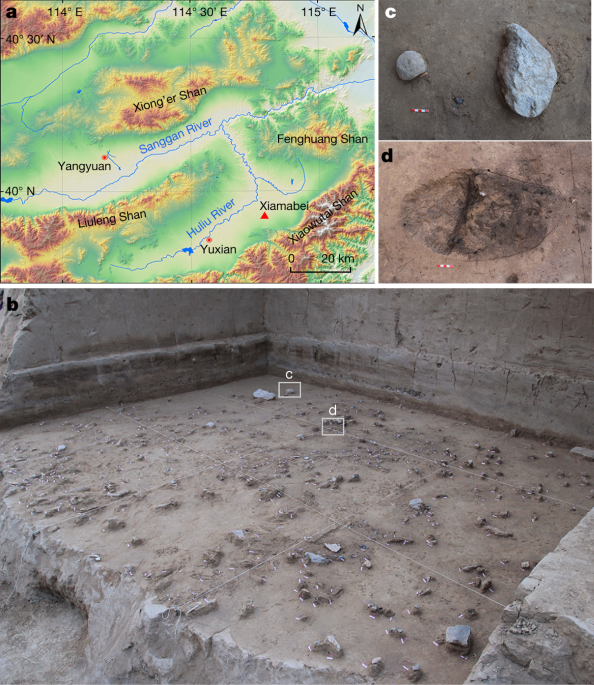
a, distribution of MIS 3 sites in northern China (see Supplementary Table A1 for site names. The source of the map https://resources.arcgis.com/). b, field view of Xiamabei. c, view of the
excavated section. d, archaeological site distributions. Key features include a red stained patch with artefacts and a hearth and surrounding ashy area. Sediment samples for composition
analysis within the excavation area are shown. Notable is the distribution of lithic artefacts and fauna, including the location of a bone tool. Stone tools submmited to use-wear analysis
provide evidence for the following activities: hafting to handles; processing of vegetal material; involvement in butchery activities and hide working; perforation of hard material; use as
wedges.
This file contains supplementary text, Figures, Tables and references.
Anyone you share the following link with will be able to read this content: