Lumbar instability remodels cartilage endplate to induce intervertebral disc degeneration by recruiting osteoclasts via hippo-ccl3 signaling

Lumbar instability remodels cartilage endplate to induce intervertebral disc degeneration by recruiting osteoclasts via hippo-ccl3 signaling


Play all audios:

Loading...

ABSTRACT Degenerated endplate appears with cheese-like morphology and sensory innervation, contributing to low back pain and subsequently inducing intervertebral disc degeneration in the


aged population.1 However, the origin and development mechanism of the cheese-like morphology remain unclear. Here in this study, we report lumbar instability induced cartilage endplate


remodeling is responsible for this pathological change. Transcriptome sequencing of the endplate chondrocytes under abnormal stress revealed that the Hippo signaling was key for this


process. Activation of Hippo signaling or knockout of the key gene Yap1 in the cartilage endplate severed the cheese-like morphological change and disc degeneration after lumbar spine


instability (LSI) surgery, while blocking the Hippo signaling reversed this process. Meanwhile, transcriptome sequencing data also showed osteoclast differentiation related gene set


expression was up regulated in the endplate chondrocytes under abnormal mechanical stress, which was activated after the Hippo signaling. Among the discovered osteoclast differentiation gene


set, CCL3 was found to be largely released from the chondrocytes under abnormal stress, which functioned to recruit and promote osteoclasts formation for cartilage endplate remodeling.


Over-expression of Yap1 inhibited CCL3 transcription by blocking its promoter, which then reversed the endplate from remodeling to the cheese-like morphology. Finally, LSI-induced cartilage


endplate remodeling was successfully rescued by local injection of an AAV5 wrapped Yap1 over-expression plasmid at the site. These findings suggest that the Hippo signaling induced


osteoclast gene set activation in the cartilage endplate is a potential new target for the management of instability induced low back pain and lumbar degeneration. SIMILAR CONTENT BEING


VIEWED BY OTHERS OSTEOPONTIN DEFICIENCY PROMOTES CARTILAGINOUS ENDPLATE DEGENERATION BY ENHANCING THE NF-ΚB SIGNALING TO RECRUIT MACROPHAGES AND ACTIVATE THE NLRP3 INFLAMMASOME Article Open


access 06 September 2024 A NEW PERSPECTIVE ON INTERVERTEBRAL DISC CALCIFICATION—FROM BENCH TO BEDSIDE Article Open access 22 January 2024 TREE SHREW AS A NEW ANIMAL MODEL FOR MUSCULOSKELETAL


DISORDERS AND AGING Article Open access 02 January 2025 INTRODUCTION Lumbar degeneration is a common disorder of the spine in the aged population, it is usually a source of low back pain.2


and a cause of disability worldwide.3 The prevalence of lumbar degeneration dramatically increases with age. Greater than 90% of people over the age of 50 present with lumbar


degeneration.4,5 The risk factors that may cause this pathology include mechanical trauma, genetic predispositions, unhealthy lifestyles and certain metabolic disorders.6,7,8 However, the


mechanism that initiates this process is not clear at present. The intervertebral disc is the largest avascular structure in the body and cells within the disc rely on diffusive transport


via the vertebral cartilage endplate (CEP) to receive nutrients, eliminate waste products and maintain disc homeostasis.9 CEP is a hyaline cartilage located on the upper and lower sides of


the intervertebral disc. Biomechanically, the endplate is known as the center of stress concentration.10,11 Despite the crucial roles of the CEP in nutrition and biomechanical stability,


vertebral endplates are extremely susceptible to mechanical failure. CEP degeneration, along with subchondral bone marrow changes were originally noticed on magnetic resonance imaging, which


is known as Modic changes (MCs).12,13 At this time, the CEP remodeling is commonly observed to occur. CEP remodeling leads to ossification with abundant pores as a cheese like morphology,


which we named the “cheese-endplate”. The cheese-endplate will lead to the difficulties in nutrient dispersion and the imbalances of disc homeostasis.14 Studies exploring the events leading


to disc degeneration have shown that failure often begins at the endplates15 and the mechanisms driving CEP transition have not been elucidated. Since CEP is the center of stress


concentration and excessive load is observed to appear on the CEP,16 studies believed the etiology of the degenerative changes could be due to mechanical micro-insults or damage secondary to


macro-insults.17 In clinic, it is observed that patients with CEP ossification and degeneration are usually accompanied by significant lumbar instability. Nowadays, the primary clinical


treatment for these conditions is to reconstruct lumbar stability with girth or internal fixation. Thus, if we can determine the underlying biological mechanism to explain CEP degeneration


that caused by lumbar instability, we would be able to identify an effective coping method to replace surgical approach for lumbar stability reconstruction, which is the major goal for this


study. Moreover, we consider that “cheese-endplate” is more than just an image finding in intervertebral disc degeneration (IVDD) patients, but rather represent an underlying pathology that


should be a target for therapy.18 Therefore, we tried to further explore the biological mechanism of disc degeneration caused by instability to demonstrate whether the lumbar endplate


degeneration caused by instability will be delayed or treated through biological targeted therapy, which will provide new ideas for clinical diagnosis and management of lumbar degeneration.


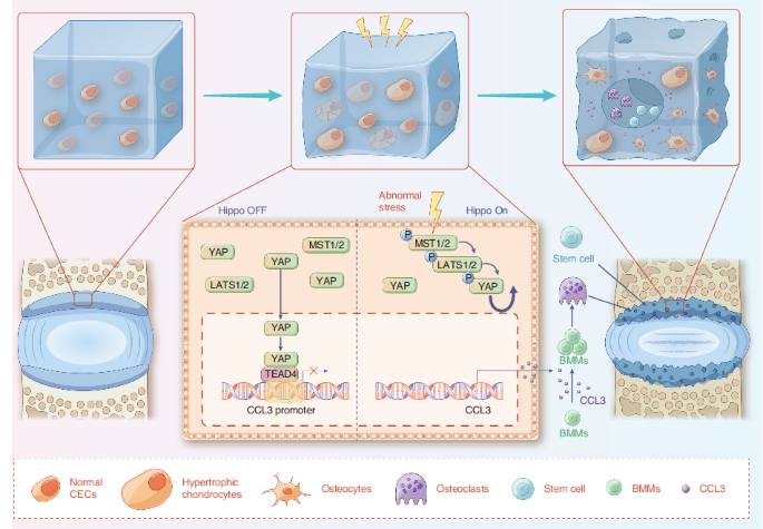
Cartilage endplate chondrocytes (CEPCs) are the cells embedded in the CEP,19,20 which are indispensable in maintaining the integrity of the structure and function of CEP.21,22,23 In this


study, we found CEPCs hold the ability to sense mechanical signals through which the abnormal stress activates Hippo signaling within the CEPCs. In addition, it is well understood that


Yes-associated protein (YAP), a key factor within the Hippo signaling, plays as a mechanosensitive factor that transmits the local mechanical cues to the nucleus and tune cellular


homeostasis.24 Recent research has confirmed that the Hippo signaling integrates diverse biochemical and mechanical cell-cell and cell-extracellular matrix signals, in that way to regulate


cell proliferation and apoptosis.25 At the same time, it shows that Yap1, the coding gene of YAP, differentially regulates chondrocyte differentiation, which promotes chondrocyte


proliferation early but inhibits subsequent maturation during skeletal development.26 However, the role of Hippo signaling in lumbar spine instability (LSI) induced endplate degeneration has


not been investigated. In this study, we investigated the role of lumbar instability induced abnormal stress in turning on the Hippo signaling that initiates cartilage endplate remodeling


through activation of osteoclast genes. Our study demonstrated that YAP is the guard for the endplate cartilage that maintains CEP homeostasis. Loss of YAP increased CCL3 release from the


CEP chondrocytes, which recruits and promotes osteoclast differentiation for cartilage endplate remodeling. Finally, we demonstrated that inhibition of Hippo signaling activation or


re-supply YAP in the CEP effectively rescued it from transforming into a cheese-like endplate and prevented the subsequent lumbar degeneration. RESULTS RADIOGRAPHY AND HISTOMORPHOLOGY OF CEP


IN THE IVDD PATIENTS AND LSI MODEL In clinic, cartilage endplate remodeling was found to occur in patients with lumbar spine degeneration. Radiological images showed these patients


presented severe ossified endplate with pores similar to that in the cheese (Fig. 1a). To better understand this pathology, we collected clinical samples of CEP from the patients with lumbar


instability. Histological staining showed the CEP was ossified as Collagen II expression decreased, accompanied with Collagen X and Osteocalcin expression increased (Fig. 1b). In order to


investigate the mechanism of this pathological change, lumbar spine instability (LSI) model was established on mice for the following test (Fig. 1c).1,27 Three-dimensional observations and


quantified analysis by Micro-CT (Fig. 1d) revealed significant changes in bone morphometry of the vertebral endplate at 8 weeks after surgery. Meanwhile, the Micro-CT analysis demonstrated


that the porosity and trabecular separation (Tb. Sp) of L5 vertebral CEP increased significantly in LSI-8 weeks (8w) mice relative to the sham group. Concomitant with the gross observation


of the degenerated disc, histological staining further revealed that the endplate was highly porotic. As shown by the Safranin O and fast green (SOFG) staining (Fig. 1e), it was observed the


CEP region in the sham group remained intact, while it became porotic and ossified in the LSI group. The green-stained bone matrix was completely surrounded the cavities, suggesting the


interfacial soft tissue was remodeled after LSI surgery. We used endplate score to visualize these pathological changes in the CEP of LSI mice, which was significantly higher than that in


the sham group (Fig. 1f). To further elucidate the remodeling process of CEP, the endplates of mice after LSI surgery for 8 weeks were subjected for immunofluorescent (IF) staining with


Collagen II and Collagen X (Fig. 1g, h). The results showed the Collagen II expression decreased significantly, while the Collagen X started to show up in the CEP of mice from the LSI group,


indicating the chondrocyte hypertrophy and cartilage calcification processes were initiated. Meanwhile, the inflammatory factor TNF-α (Fig S1A) and apoptotic marker TUNEL (Fig S1B), along


with the expression of vascular-related marker CD31 (Fig S1C) and the nerve-related marker CGRP (Fig S1D), in the CEP pores further supported our observation of cartilage endplate remodeling


in the LSI model. The above results demonstrate LSI induced abnormal stress initiates CEP remodeling to a cheese-like ossified tissue. FINITE ELEMENT ANALYSIS (FEA) SIMULATED CEP MECHANICAL


STRESS DISTRIBUTION AFTER LSI SURGERY To understand the mechanical stress changes in the endplates of mice in lumbar instability conditions, we performed computerized finite element


analysis (FEA) to map the mechanical stress distribution in the endplates of mice with sham or LSI surgery. We first assigned material properties (Table S1) to each part of the model in


Hypermesh software as described in the literature.28,29,30,31 (Fig. 2a and Fig S2). We then applied forces to the spine unit to mimic the upright, lateral bending and rotation positions of


the lumbar spine (Fig. 2b). As a result, the maximum principal strain in LSI mice were markedly elevated in the whole CEP region, in which the posterior part showed the most significant


stress change (Fig. 2c). Together, our findings indicate that the mechanical stress distribution in CEP was altered after the LSI surgery, giving the rationale that CEP undergoes remodeling


after sensing the changes of mechanical stimulation. ABNORMAL STRESS INDUCES CEP CHONDROCYTES DEGENERATION In order to simulate the mechanical stress of CEP chondrocytes (CEPCs) in vitro, a


loaded cell culture system was used to apply various levels of mechanical stress, which is represented by cyclic tensile strain (CTS).32 The endplate chondrocytes were isolated and cultured


on the system. To understand how the abnormal mechanical stress initiates CEP remodeling, we firstly determined the exact range and frequency of the tensile equipment to mimic the


degenerative conditions of CEPCs in vivo. In the control group, no stress was applied, while 5% and 12% range with 0.5 Hz were applied in the other two groups.32 Each group was stretched for


8 h daily, and samples were collected after seven days (Fig. 3a). Then, the CEPCs from the three groups were subjected to IF staining, showing gradually decreased Collagen II expression


accompanied with increased Collagen X expression from the control group to the 12% range group (Fig. 3b, c). We further stained MMP3 and Ki67, a marker of cell proliferation. The results


confirmed that MMP3 expression was increased while Ki67 expression was decreased under abnormal stress (Fig S3). The RT-qPCR (Fig. 3d) and Western blot (Fig. 3e) analysis showed similar


trend. These results suggest the 12% range group of cells under CTS were transforming toward hypertrophic chondrocytes, which was comparable to the change of CEPCs after LSI surgery in vivo.


Based on the data above, we used 12% CTS treated CEPCs as the in vitro abnormal mechanical simulation model for the following experiment. ABNORMAL STRESS REGULATES THE HIPPO SIGNALING IN


CEPCS To elucidate how mechanical stress initiates the CEP remodeling, we performed transcriptome analysis of the CEPCs from the control and 12% range groups. The results showed 722


differentially expressed genes (DEGs) were down-regulated and 1 290 DEGs were up-regulated. KEGG analysis with the above DEGs (Fig. 4a) revealed the Hippo signaling was significantly


enriched in the down-regulated DEGs, while the osteoclast differentiation pathway, on the contrary, was enriched in the up-regulated pool of DEGs. Furthermore, gene set enrichment analysis


(GSEA, Fig. 4b) also showed clearly that the Hippo signaling was significantly changed after 12% CTS, with the trend of down-regulation. As proposed by the transcriptome analysis, we then


examined the endogenous expression of YAP, a key mediator of Hippo signaling, in the CEP from mice underwent LSI surgery or sham surgery for 8 weeks. As expected, strong YAP expression was


observed in all the CEP areas in the sham group, while it was remarkably decreased in the LSI group (Fig. 4c). These data suggest a reduction of YAP in chondrocytes under abnormal mechanical


stress. IF staining further verified the above results by showing that 5% range of mechanical stress slightly increased the YAP expression in the nucleus, but 12% range of mechanical stress


blocked it from entering the nucleus of the endplate chondrocytes (Fig. 4d–g). Western blot analysis also showed that 12% range of mechanical stress reduced YAP and increased its


phosphorylation (p-YAP) level (Fig. 4h). In parallel experiments, 5% CTS slightly increased the YAP expression and narrowed the p-YAP/YAP ratio. Altogether, these findings suggest that the


Hippo signaling status and the YAP expression are highly correlated to the mechanical stress loaded on the endplate chondrocytes. YAP1 DELETION DETERIORATES CEP REMODELING AFTER LSI SURGERY


To investigate whether YAP is required to protect the integrity of CEP cartilage, we first labeled the CEP resided chondrocytes with Col2a1 in red by crossing the _Col2a1-CreER_ mice with


TdTomato mice (_Col2a1-ER__Tdt_) (Fig. 5a). Then, we performed IF staining of YAP on CEP. The results showed that YAP was highly expressed and merged with Col2a1+ chondrocytes in CEP (Fig.


5b), suggesting abundant expression of YAP in CEP under physical conditions. Next, we genetically removed YAP in chondrocytes by generating _Col2a1-CreER::Yap1__fl/fl_ (_Yap1__fl/fl,Col2a1_)


mice. The knockout efficiency was confirmed by immunohistochemical staining of YAP, showing substantial reduction of YAP expression from CEP in the _Yap1__fl/fl,Col2a1_ mice as compared to


the wild type (WT, _Yap1__fl/fl_) ones (Fig. 5c). As observed, no obvious cartilage defect was observed in the _Yap1__fl/fl,Col2a1_ mice after sham surgery, while the CEP cartilage


degradation was more severe in the _Yap1__fl/fl,Col2a1_ mice than that of the control mice after LSI surgery (Fig. 5d, e). SOFG staining showed the cartilage area had been completely


replaced by bone tissue and the cavities were fused to became larger, indicating the cartilage remodeling was stronger in the _Yap1__fl/fl,Col2a1_ mice (Fig. 5f), which was further confirmed


by the endplate score evaluation (Fig. 5g). To further explain the remodeling process, we applied IF staining of Collagen II, Collagen X and TNF-α in the CEP tissue (Fig. 5h–j).


Quantitative analysis showed the expression of Collagen II was significantly reduced but Collagen X and TNF-α was significantly increased in the CEP after LSI surgery in both the WT and


_Yap1__fl/fl,Col2a1_, among which, the expression change of these markers were more significant in the _Yap1__fl/fl,Col2a1_ group (Fig. 5k–m). The results above demonstrate YAP is necessary


to protect CEP cartilage from remodeling and degeneration under abnormal stress stimulation. ACTIVATION OF YAP ATTENUATES CEP REMODELING IN LSI MICE As we found that Hippo signaling mediates


abnormal stress inudced CEP degeneration, then we decided to further explore its function in initiating the CEP remodeling. First, we used pharmacological treatment with inhibitors that


regulate Hippo signaling. When the Hippo signaling is activated, a series of phosphorylation events via MST and LATS kinases ultimately leads to the phosphorylation of YAP. Since central to


the Hippo signaling is a kinase cascade consisting of MST1/2 and LATS1/2, we used Lats-IN-1, an inhibitor to LATS1/2 thereby activating YAP,33 in the following study. Wild-type mice treated


daily with Lats-IN-1 showed a marked resistance to abnormal stress induced CEP degradation compared with controls (Fig. 6a). Lats-IN-1 treated mice showed less pores or remodeling related


signs in the CEP (Fig. 6b, c), with abundant Collagen II expression and rare Collagen X expression (Fig. 6d). Moreover, Lats-IN-1 was also applied to the CEPCs in vitro, and all of IF


staining, Western blot and RT-qPCR showed that Lats-IN-1 prevented the process of CEP from degeneration (Fig. 6e–h). These data demonstrated that pharmacological induction of YAP protects


CEP cartilage from degeneration. ABNORMAL STRESS ALLOWS OSTEOCLAST RECRUITMENT IN CEP TO INITIATE REMODELING THROUGH THE HIPPO SIGNALING Since the pores and remodeling of the CEP were


observed in the LSI mice, it gives the possibility on the activation of osteoclasts under the condition of lumbar instability. As described above, the transcriptome data pointed out the


osteoclast differentiation signaling was significantly enriched based on the DEGs screened from the 12% CTS versus the control, which means an osteoclast related gene set was up regulated


during this process. To confirm these, we combined the heatmap of the DEGs that related to osteoclast (Fig. 7a) and GESA analysis (Fig. 7b) together. It showed that the genes for osteoclast


recruitment and formation were largely activated in the CEPCs after 12% CTS treatment. As expected, TRAP staining presented that osteoclasts were increased in the _Yap1__fl/fl,Col2a1_ mice


CEP with LSI surgery (Fig. 7c, d), which was in accordance with the observed cavities enlargement in the CEP, indicating a more active remodeling process was occurred. To further confirm


this, we used _Ctsk-Cre::TdTomato_ (_Ctsk__Tdt_) mice to label osteoclasts, which were then performed with LSI surgery. The harvested CEP was co-stained with osteocalcin. It presented that


the osteoclasts and osteoblasts were both actively showed up in the cavities of the CEP under the same time-spatial condition, indicating the remodeling in CEP was indeed processed when


abnormal stress was applied (Fig. 7e). Furthermore, Nestin-1 positive stem cells were found in the cavities of the CEP, further suggesting the stem cells were recruited to the remodeling


site for this process (Fig. 7e). However, we did not change the expression of YAP in osteoclasts directly, so the activation of osteoclasts should be due to the indirect effect from the


CEPCs under abnormal stress condition. To confirm this hypothesis, we first collected the supernatant of the CEPCs from the cells with 12% CTS treatment, and then applied it for osteoclast


induction. We observed that the cells in the 12% CTS supernatant group became osteoclastogenesis earlier and stronger than the control group (Fig. 7f). RT-qPCR analysis also suggested


similar results by showing increased expression of osteoclast-activating genes (Fig. 7g). Therefore, we conclude that 12% CTS induces CEPCs to release cytokines to recruit and promote


osteoclast formation in the CEP to start remodeling under abnormal stress. LOSS OF YAP1 FACILITATES CEPCS TO RELEASE CCL3 FOR OSTEOCLAST RECRUITMENT AND FORMATION By reviewing the


transcriptome data, we found that chemotactic cytokine ligand 3, CCL3 (MIP-1α, macrophage inflammatory protein-1α) was highly expressed in the CEPCs after 12% CTS treatment (Fig. 8a). Elisa


evaluation of the CCL3 in the supernatant was performed and the results showed a two-fold increase in the 12% CTS medium, suggesting 12% CTS induced CCL3 generation and release in CEPCs


(Fig. 8b). Then, immunohistochemical staining confirmed that CCL3 was significantly expressed during CEP remodeling in vivo (Fig. 8c). The extracted protein was evaluated by western blot and


the results showed the same trend (Fig. 8d). To further confirm the function of CCL3 in osteoclast precursors-bone marrow monocytes (BMMs) recruitment, transwell assay was performed. It


showed that more BMMs migrated to the lower chamber with CCL3 added into the medium when compared with the blank (Fig. 8e). The results above demonstrate that the instability induced


abnormal stress initiates osteoclasts recruitment and formation by CEPCs releasing CCL3. To further explore the CCL3 interaction with Yap1 in abnormal stress induced CEP remodeling, we


firstly evaluated the YAP and CCL3 expression in 12% CTS treated or Lats-IN-1 treated or both treated CEPCs with Western blot and IF staining. As expected, the CCL3 expression was increased


under abnormal stress, however, it was suppressed when YAP was activated in CEPCs (Fig. 8f, g), indicating that YAP is a regulator to control CCL3 expression. Meanwhile, an anti-CCL3


antibody as a neutralizing antibody was added into the supernatant of 12% CTS treated CEPCs, which was then applied to induce osteoclast. The results reflected that the anti-CCL3 antibody


significantly inhibited osteoclast activation in a dose dependent manner (Fig. 8h, i), suggesting the CCL3 release from the CEPCs under abnormal stress was a key factor in activating


osteoclastogenesis. To explore the undermined molecular mechanism, we tested the effect of YAP on different CCL3 promoter regions. Generally, YAP-overexpression inhibited CCL3-luciferase


reporter activity as compared to the control, which was more significant in the -1 043 to +76 promoter region (Fig. 8j, k). As YAP has no direct binding sites on the promoter, it was


speculated that its function should be exerted by forming the complex with TEADs. Then, we evaluated different TEADs expression in the CEPCs from the 12% CTS and control groups by RT-qPCR.


The data showed the change of TEAD4 was similar like YAP under abnormal stress (Fig. 8l), indicating the YAP/TEAD4 transcriptional complex might possibly regulate the expression of CCL3. The


above data suggest that YAP degradation unlocked CCL3 expression in CEPCs, which is then released to mediate osteoclast activity in CEP under abnormal stress condition. YAP1-AAV5 INJECTION


SUPPRESSES CEP REMODELING TO RESCUE LSI INDUCED CHEESE-LIKE ENDPLATE The above experiments suggested that endogenous YAP plays a key role in maintaining CEP tissue homeostasis, which could


be a potential target to rescue CEP from remodeling. Then, we chose adeno-associated virus serotype 5 (AAV5) as a carrier34 to wrap the YAP over-expression plasmid, naming Yap1-AAV5. It was


injected into the left part of caudal endplate in the level of L4-5 in mice (Fig. 9a). Firstly, to confirm the successful transfection of Yap1 over-expression plasmid with this approach, we


evaluated the green fluorescence in the CEP. By co-staining with YAP, it showed clearly that YAP was largely overlapped with the green fluorescence from the plasmid, indicating this


injection method worked well to drive potent and long-lasting YAP expression in the CEP after injection for eight weeks (Fig. 9b). Next, we applied Yap1-AAV5 in conjunction with LSI surgery


or sham in the wild type mice. Micro-CT showed that Yap1-AAV5 injection well prevented CEP from degeneration after LSI surgery (Fig. 9c–e). As shown by SOFG staining, no obvious ossified


tissue was observed in the CEP of Yap1-AAV5 treated mice as well (Fig. 9d, e). The down-stream rescuing effect was further verified by IHC staining of Collagen II, Collagen X and CCL3. The


Collagen II expression, which was found to remain at a high level at 8 weeks after LSI and Yap1-AAV5 injection, and Collagen X and CCL3 expression in the CEP with LSI and Yap1-AAV5 treatment


was kept in a low level (Fig. 9h–j). Taken together, our results demonstrate that Yap1 is an effective therapeutic target to prevent abnormal stress induced CEP remodeling, and the


Yap1-AAV5 injection is an ideal approach to manage CEP from degradation under abnormal stress conditions. DISCUSSION Current clinical treatments for lumbar degeneration include


pharmacological and surgical interventions, but which lack the ability to halt disease progression and restore intervertebral disc function.35 Abundant research work is mainly focusing on


regenerating degenerated intervertebral discs, including usage of growth factors and stem cell transplantation etc.36,37 However, studies of this kind are still in their infancy. Therefore,


understanding the key mechanisms of lumbar degeneration is beneficial for us to find therapeutic targets. Since lumbar degeneration usually begins with aging or mechanical trauma, these


allow the endplate to undergo sclerosis and become porous, which is clinically associated with low back pain.18,38 In addition, several studies have confirmed that a series of remodeling


changes occur in the endplate during this process.39 However, the connection between abnormal stress and cheese-endplate is not well explained. Our study shows that lumbar instability


induces abnormal endplate stress, which activates Hippo signaling in CEPCs, leading to lumbar degeneration. Chondrocytes can sense the magnitude or type of mechanical stimuli in the


environment and respond accordingly.40 Normally, an appropriate mechanical load generated by body weight and muscle strength is necessary to maintain bone and cartilage homeostasis.41 At the


same time, studies have shown that mechanical stress is an important cause of articular cartilage degeneration.42,43 Researchers suggest that mechanical injury predisposes the metabolic


balance of chondrocytes to be disturbed, leading to cartilage degradation development, and it is believed to be the most critical etiologic factors.44,45 Non-physiological mechanical


stimulation inhibits collagen synthesis and promotes its degradation in cartilage,46 which may cause an inflammatory response in cartilage and aggravate the degradative phenotype. The


current study focused on inhibiting these inflammatory signals47 to counteract the inflammatory damage caused by abnormal stress and alleviate the degenerative phenotype. Degradation of


collagen in cartilage due to mechanical damage, aging and other objective causes impairs the ability of cartilage to load stress.48 Without changing the abnormal stress itself, we aim to


find the key to explain cartilage degeneration caused by abnormal stress and the subsequent bad effects of stress more fundamentally from biological view, thereby conferring protection


strategies to cartilage structure and functionality. YAP and TAZ are the nuclear transducers of the Hippo pathway.49 Dupont et al. identified YAP/TAZ as sensors and mediators of mechanical


cues instructed by the cellular microenvironment.24 Deng et al. established a reciprocal antagonism between Hippo-YAP/TAZ and NF-κB signaling in regulating the induction of matrix-degrading


enzyme expression and cartilage degradation during osteoarthritis pathogenesis.50 Our findings suggest that loss of Yap1 in chondrocytes accelerates CEP degradation and initiates its


remodeling. In contrast, treatment with YAP agonist inhibited endplate remodeling. Meanwhile, in this study, we found that YAP mediates the function of Hippo signaling to control CEP


homeostasis during LSI by antagonizing CCL3 release. Upon excessive mechanical stress, the inflammatory cytokine CCL3, originally inhibited by YAP, recruits and activates osteoclasts.


Previous studies have established that osteoclast lineage cells instigate porosity of sclerotic endplates and spinal pain behavior. Osteoclastogenic chemokine CCL3 has been demonstrated to


enhance RANKL-induced OC formation51,52 and to regulate macrophage trafficking to the inflamed joint.53 We found that CCL3 is regulated by Yap1 and is abnormally elevated under abnormal


stress to accelerate CEP remodeling. Finally, adeno-associated virus (AAV) serotype 5 was used as the vector.54 Huang et al. used intraarticular administration of AAV5, by which effective


and durable expression in cartilage was still detected up to three months.34 In the present study, Yap1-AAV5 was used to overexpress Yap1 in CEP and successfully prevented CEP degeneration


induced by abnormal stress stimulation in mice. Remarkably, experimental activation of YAP/TAZ in mice can promote regeneration in organs with poor or compromised regenerative capacity.55


However, therapeutic YAP/TAZ activation may cause serious side effects. Notably, YAP/TAZ are hyperactivated in human cancers, and prolonged activation of YAP/TAZ triggers cancer


development.56,57 Therefore, the local application effect is more valuable for clinical transformation. In conclusion, our study revealed the abnormal mechanical stress changes the Hippo


signaling and reduces the expression of YAP, which losses its suppression function on cytokine CCL3 to recruit and activate osteoclasts for CEP remodeling. Our study suggests that YAP is


essential for maintaining a healthy CEP, as well as the intervertebral disc. MATERIALS AND METHODS HUMAN SUBJECTS All human endplate samples and imaging data were obtained from the surgical


patients, and the informed consent of the patients had been obtained in advance, which was reviewed and approved by the Ethics Committee of the First Affiliated Hospital of Soochow


Unviersity. MICE Heterozygous _Col2a1-CreER_ mice were crossed with _Yap1__flox/flox_ mice. The offspring were intercrossed to generate _Col2a1-CreER::YAP__flox/flox_ (conditional deletion


of Yap1 in Col2a1 lineage cells, referred to as _Yap1__fl/fl,Col2a1_ herein). The _Col2a1-CreER_ mouse strain needs tamoxifen injection. Briefly, as we did in our previous paper,58 mice were


intraperitoneally injected with tamoxifen (T5648, Sigma-Aldrich) at a concentration of 10 mg/mL for five consecutive days. The genotypes of the mice were determined by PCR analyses of


genomic DNA, which was extracted from mouse tails with the following primers: _Col2a1-CreER:_ forward: 5ʹ- GATCTCCGGTATTGAAACTCCAGC -3ʹ, reverse: 5ʹ- GCTAAACATGCTTCATCGTCGG-3ʹ; _Ctsk-cre:_


forward: 5ʹ- GATCTCCGGTATTGAAACTCCAGC -3ʹ, reverse: 5ʹ- GCTAAACATGCTTCATCGTCGG -3ʹ; YAP loxP allele forward: 5ʹ- GGCACTGTCAATTAATGGGC-3ʹ, reverse: 5ʹ-AGTCTGTAACAACCAGTCAGGGA -3ʹ, WT: 5ʹ-


TCCATTTGTCCTCATCTCTTACTAAC-3ʹ; TdTomato loxP allele _tdt1:_ forward: 5ʹ-AAGGGAGCTGCAGTGGAGTA-3ʹ, tdt1: forward: 5ʹ-AAGGGAGCTGCAGTGGAGTA-3ʹ, _tdt2:_ forward: 5ʹ-CCGAAAATCTGTGGGAAGTC-3ʹ,


_tdt3:_ forward: 5ʹ- GGCATTAAAGCAGCGTATCC-3ʹ, _tdt4:_ forward: 5ʹ- CTGTTCCTGTACGGCATGG-3ʹ. LSI surgery was performed using 8-week-old male mice. Briefly, in order to create lumbar


instability, the spinous processes, supraspinous and interspinous ligaments of the L3-L5 were all excised.1,59 In sham operation, only L3-5 paraspinal muscles were dissected. Samples were


collected eight weeks after surgery. In the drug treatment portion, Lats-IN-1 mice received daily intraperitoneal injections of 3 mg/kg.33,60 The control group used the same amount of


dimethyl sulfoxide (DMSO). Samples were collected after eight consecutive weeks. The performance of the animals in this study was approved by the Ethics Committee of the First Affiliated


Hospital of Soochow Unviersity. MICRO-CT The whole lumbar spine was dissected and evaluated with micro-CT (SkyScan 1176, SkyScan, Aartselaar, Belgium) with the following settings, namely, 65


 kV, 385 mA, and 0.5 mm Al filter. Three-dimensional (3D) reconstruction was performed with system software. Coronal images of the L4–L5 unit were used to perform three dimensional


histomorphometric analyses of the caudal endplate. Three-dimensional structural parameters analyzed were total porosity and Tb.Sp for the endplates. Five consecutive coronal-oriented images


were used for showing 3-dimensional reconstruction of the endplates. HISTOMORPHOLOGICAL AND IMMUNOHISTOCHEMICAL ANALYSIS The harvested specimens were fixed for 24-48 h and then decalcified


for 2 weeks at room temperature. Following that, the spine was embedded in paraffin waxor optimal cutting temperature (OCT) compound (Sakura Finetek, Torrance, CA). Five-μm-thick coronal


sections of the L4–L5 lumbar spine were processed for SOFG, TRAP (Sigma-Aldrich), and immunohistochemistry (IHC) staining. Ten-μm-thick coronal frozen sections were prepared for


immunofluorescent staining. The primary antibodies used for staining included Collagen II (1:200, ab34712, Abcam), Collagen X (1:200, ab260040, Abcam), YAP1 (1:200, ab205270, Abcam), CCL3


(1:400, ab179638, Abcam), Nestin (1:100, ab221660, Abcam), TNF-α (1:200, ab183218, Abcam), CD31 (1:200, ab76533, Abcam), CGRP (1:50, ab81887, Abcam), Osteocalcin (1:200, M173, TAKARA), CCL3


(10 μg/mL, AF-450-NA, R&D system). Images were obtained using a microscope (Zeiss Axiovert 200; Carl Zeiss Inc., Thornwood, NY). ImageJ (NIH) software was used for quantitative analysis.


We calculated endplate scores as described previously.61,62 TUNEL ASSAY Frozen sections were stained with a One Step TUNEL Apoptosis Assay Kit (C1086, Beyotime) according to manufacturer’s


protocol. The images were captured with a fluorescence microscope (Zeiss Axiovert 200). FINITE ELEMENT ANALYSIS Lumbar finite element model (L4‑L5). In an FEM, a geometrical complex spine


segment can be divided into different regions according to its anatomical structure, and then meshed with various types of elements. Each region can be assigned an appropriate material model


to reflect its biomechanical characteristics. In the present study, lumbar Micro-CT scan images of a normal 8 weeks-male-C57BL6/J mouse were used. Then the solid model was constructed in


HyperMesh (Altair Engineering, Inc., Troy, MI, USA) and the material properties of each part of the model were assigned according to the description in the literature.29,30,31 (Table S1).


Finally, biomechanical finite element analysis of the L4‑L5 segment was carried out in ABAQUS/Standard (Dassault Systemes, Velizy ‑Villacoublay, France). The model included the vertebrae,


intervertebral discs, endplates and ligaments. ISOLATION OF CARTILAGE ENDPLATE CHONDROCYTES Primary CEPCs were isolated from the 4-6 week mouse. Specifically, CEP tissues of mouse lumbar


spine were trimmed with sharp knife tips. After collecting lumbar endplate cartilage from 10 to 15 mice at a time, the cartilage was washed more than three times using sterile PBS. After 20 


min of Trypsin (Gibco) digestion, the plates were again washed three times with sterile PBS, The CEP tissues were then changed to Collagenase (C6885, Sigma-Aldrich 0.2 mg/mL) solution in


serum-free medium. The CEP tissue was cut as finely as possible in solution and blown every half hour during digestion. After 5–6 h, the cell suspension was resuspended by centrifugation and


with F12 medium (10% FBS) in a dish culture completely, once every two days in liquid. The extracted cells were identified by immunofluorescence staining, and the results confirmed that the


extracted cells expressed a large amount of Collagen II protein. MECHANICAL LOADING A Loaded Cell Culture System (Celload-300) was used to apply mechanical stimulation on CEPCs. CEPCs were


seeded on a stretched die of polydimethylsiloxane (PDMS, Dow Corning) at an initial density of 10 000 cells/cm2 In the stretching group, CEPCs were subjected at a tensile strength of 5% or


12% at 0.5 Hz for 8 h per day for 7 consecutive days. YAP agonists Lats-IN-1 (10 μmol/L, MCE) and inhibitor Verteporfin (2 μmol/L, Sigma-Aldrich) was supplemented in culture medium during


the entire course of mechanical loading.33,63,64 IMMUNOFLUORESCENCE STAINING OF CEPCS Cells were fixed with 4% PFA for 20 min after mechanical treatment, followed by staining according to


conventional methods. Briefly, the sections were incubated with primary antibodies to Collagen II (1:200, ab34712, Abcam), Collagen X (1:200, ab260040, Abcam), YAP1 (1:200, ab205270, Abcam),


phosphor-YAP1 (1:200, ab76252, Abcam), Osteocalcin (1:200, M173, TAKARA), CCL3 (1:200, ab179638, Abcam). The images were observed and captured by a fluorescence microscope (Zeiss Axiovert


200) or confocal microscope (Zeiss LSM 780). ImageJ (NIH) software was used for quantitative analysis. WESTERN BLOT (WB) WB analysis was conducted on the protein lysates from the


mechanically and drug-treated cells. Specific antibodies were applied for incubation, and the proteins were detected by using an ECL Western Blotting Substrate Kit (ab65623, Abcam). The


antibodies used for WB were Collagen II (1:1 000, AF0135, Affinity), Collagen X (1:1 000, ab182563, Abcam), Osteocalcin (1:500, sc-365797, Santa Cruz), YAP (1:1 000, 14074, Cell Signaling


Technology), Phospho-YAP (1:1 000, 13008, Cell Signaling Technology), CCL3 (1:1 000, ab179638, Abcam), and GAPDH (1:5 000, ab8245, Abcam). RT-QPCR The total RNA was extracted from lumbar


spinal endplate tissue samples using TRIzol reagent (Invitrogen). RNA was reverse transcribed into complementary DNA using the All-In-One System (Abm). RT-qPCR was performed with Supermix


(Bio-Rad Laboratories) on a C1000 Thermal Cycler (Bio-Rad Laboratories). Relative expression was calculated for each gene by the 2−ΔΔCT method, with glyceraldehyde 3-phosphate dehydrogenase


(GAPDH) for normalization. The primers used for RT-qPCR are listed in Table S2. TRANSCRIPTOME ANALYSIS CEPCs treated with different CTS were analyzed. The transcriptome sequencing was


conducted by OE biotech Co. Ltd. (Shanghai) Cleaning reads were obtained using Trimmomatic and mapped to reference genome using hisat2. FPKM (fragments per kilobase of exon per million reads


mapped) value of each gene was calculated using cufflinks. The DEGs, GESA (Gene Set Enrichment Analysis), and KEGG (Kyoto Encyclopedia of Genes and Genomes) enrichment analysis were


performed using R software. _P_ < 0.05 and FoldChange > 2 or FoldChange < 0.5 was set as the threshold for significantly differential expression or differential enrichment.The venn


and volcano plot analysis of DEGs were performed using the OE suite of online tools (https://cloud.oebiotech.cn/task/). The clustered heatmap in Fig. 7A was plotted using R package


‘pheatmap’ (ver. 1.0.12). OSTEOCLAST INDUCTION Primary mouse bone marrow monocytes (BMMs) were isolated from tibia and femur of 6-week-old mice. Briefly, BMMs were cultured in a-MEM medium


containing 30 ng/mL M-CSF (R&D Systems). RANKL (50 ng/mL, R&D Systems) was used to induce osteoclast differentiation. In this experiment, supernatant from mechanically treated cells


was used instead of a-MEM media. In the neutralization test, goat anti-CCL3 antibody (AF-450-NA, R&D system) or goat serum (Sigma-Aldrich) diluted in saline was used at the same IgG


concentration. TRAP staining (Sigma-Aldrich) was conducted according to the manufacturer’s instructions. ELISA The concentration of CCL3 was determined by using the Mouse MIP-1α (Macrophage


Inflammatory Protein 1 Alpha) ELISA Kit (Elabscience) according to the manufacturer’s instructions. TRANSWELL ASSAY A total of 2 × 10⁴ BMMs were placed in the upper chamber of a 24-well


transwell (Corning, NY). Then, CCL3 Protein (MedChemExpress) was dissolved in a-MEM media with different concentrations in the lower chamber. After 48 h, the cells were stained with 0.1%


crystal violet solution (Beyotime) for observation and quantification. DUAL-LUCIFERASE ASSAY The amplified promoter of Yap1 was subcloned into the firefly plasmids in the pGL3BASIC


luciferase vector. At last, a Dual-Luciferase Reporter Assay System (Promega) was used to evaluate luciferase activity. Each procedure of these experiments was repeated three times


independently. AAV INJECTION 8-week-old male C57BL/6 J mice were used to perform LSI or sham surgery (8 per group). AAV5-Yap1 was purchased from Genomeditech. We fully exposed the caudal


endplate of L4–L5 with a ventral approach. Then, 1 × 108 AAV particles in a 10 μL volume was injected into the left part of caudal endplate of L4–L5 using borosilicate glass capillaries


after drilling a hole at left part of endplate.1 Ten-μm-thick frozen sections were used, and the GFP signals were inspected under a fluorescence microscope (Zeiss Axiovert 200). STATISTICAL


ANALYSIS The comparisons between multiple groups were performed using multiple comparisons by one-way ANOVA. For RT-qPCR data expressed as relative fold changes, Student’s _t_ test and


one-way ANOVA with Dunnett’s test were used for pairwise comparisons and multi-group comparison, respectively. Results are represented as mean ± s.d. _P_ values < 0.05 were considered to


be significant. Equal variances were assumed. All analyses were performed with GraphPad Prism software (Version 7.0). No statistical methods were used to predetermine sample size. The


experiments were not randomized and the investigators were not blinded to allocation during experiments and outcome assessments. REFERENCES * Ni, S. et al. Sensory innervation in porous


endplates by Netrin-1 from osteoclasts mediates PGE2-induced spinal hypersensitivity in mice. _Nat. Commun._ 10, 5643 (2019). Article  CAS  PubMed  PubMed Central  Google Scholar  *


Andersson, G. B. Epidemiological features of chronic low-back pain. _Lancet_ 354, 581–585 (1999). Article  CAS  PubMed  Google Scholar  * Centers for Disease, C. & Prevention Prevalence


and most common causes of disability among adults–United States, 2005. _MMWR Morb. Mortal. Wkly Rep_. 58, 421–426 (2009). * Teraguchi, M. et al. Prevalence and distribution of intervertebral


disc degeneration over the entire spine in a population-based cohort: the Wakayama Spine Study. _Osteoarthr. Cartil._ 22, 104–110 (2014). Article  CAS  Google Scholar  * Cheung, K. M. et


al. Prevalence and pattern of lumbar magnetic resonance imaging changes in a population study of one thousand forty-three individuals. _Spine (Philos. Pa 1976)_ 34, 934–940 (2009). Article 


Google Scholar  * Urban, J. P. & Roberts, S. Degeneration of the intervertebral disc. _Arthritis Res. Ther._ 5, 120–130 (2003). Article  PubMed  PubMed Central  Google Scholar  * Clouet,


J. et al. The intervertebral disc: from pathophysiology to tissue engineering. _Jt. Bone Spine_ 76, 614–618 (2009). Article  Google Scholar  * Hadjipavlou, A. G., Tzermiadianos, M. N.,


Bogduk, N. & Zindrick, M. R. The pathophysiology of disc degeneration: a critical review. _J. Bone Jt. Surg. Br._ 90, 1261–1270 (2008). Article  CAS  Google Scholar  * Applebaum, A.,


Nessim, A. & Cho, W. Modic change: an emerging complication in the aging population. _Clin. Spine Surg._ 35, 12–17 (2022). Article  PubMed  Google Scholar  * Desmoulin, G. T., Pradhan,


V. & Milner, T. E. Mechanical aspects of intervertebral disc injury and implications on biomechanics. _Spine (Philos. Pa 1976)_ 45, E457–E464 (2020). Article  Google Scholar  * Adams, M.


A., Freeman, B. J., Morrison, H. P., Nelson, I. W. & Dolan, P. Mechanical initiation of intervertebral disc degeneration. _Spine (Philos. Pa 1976)_ 25, 1625–1636 (2000). Article  CAS 


Google Scholar  * de Roos, A., Kressel, H., Spritzer, C. & Dalinka, M. MR imaging of marrow changes adjacent to end plates in degenerative lumbar disk disease. _AJR Am. J. Roentgenol._


149, 531–534 (1987). Article  PubMed  Google Scholar  * Modic, M. T., Steinberg, P. M., Ross, J. S., Masaryk, T. J. & Carter, J. R. Degenerative disk disease: assessment of changes in


vertebral body marrow with MR imaging. _Radiology_ 166, 193–199 (1988). Article  CAS  PubMed  Google Scholar  * Ashinsky, B. G. et al. Intervertebral disc degeneration is associated with


aberrant endplate remodeling and reduced small molecule transport. _J. Bone Min. Res_. 35, 1572–1581 (2020). Article  Google Scholar  * Natarajan, R. N., Ke, J. H. & Andersson, G. B. A


model to study the disc degeneration process. _Spine (Philos. Pa 1976)_ 19, 259–265 (1994). Article  CAS  Google Scholar  * Smith, L. J., Nerurkar, N. L., Choi, K. S., Harfe, B. D. &


Elliott, D. M. Degeneration and regeneration of the intervertebral disc: lessons from development. _Dis. Model Mech._ 4, 31–41 (2011). Article  PubMed  Google Scholar  * Kushchayev, S. V. et


al. ABCs of the degenerative spine. _Insights Imaging_ 9, 253–274 (2018). Article  PubMed  PubMed Central  Google Scholar  * Dudli, S., Fields, A. J., Samartzis, D., Karppinen, J. &


Lotz, J. C. Pathobiology of modic changes. _Eur. Spine J._ 25, 3723–3734 (2016). Article  PubMed  Google Scholar  * He, Z., Jia, M., Yu, Y., Yuan, C. & Wang, J. Roles of SDF-1/CXCR4 axis


in cartilage endplate stem cells mediated promotion of nucleus pulposus cells proliferation. _Biochem. Biophys. Res. Commun._ 506, 94–101 (2018). Article  CAS  PubMed  Google Scholar  * He,


Z., Pu, L., Yuan, C., Jia, M. & Wang, J. Nutrition deficiency promotes apoptosis of cartilage endplate stem cells in a caspase-independent manner partially through upregulating BNIP3.


_Acta Biochim. Biophys. Sin. (Shanghai)_ 49, 25–32 (2017). Article  CAS  PubMed  Google Scholar  * Wang, H. et al. Utilization of stem cells in alginate for nucleus pulposus tissue


engineering. _Tissue Eng. Part A_ 20, 908–920 (2014). Article  CAS  PubMed  Google Scholar  * Chen, S. et al. Mesenchymal stem cells protect nucleus pulposus cells from compression-induced


apoptosis by inhibiting the mitochondrial pathway. _Stem Cells Int._ 2017, 9843120 (2017). Article  PubMed  PubMed Central  Google Scholar  * Wang, W. et al. Transplantation of


hypoxic-preconditioned bone mesenchymal stem cells retards intervertebral disc degeneration via enhancing implanted cell survival and migration in rats. _Stem Cells Int._ 2018, 7564159


(2018). Article  PubMed  PubMed Central  Google Scholar  * Dupont, S. et al. Role of YAP/TAZ in mechanotransduction. _Nature_ 474, 179–183 (2011). Article  CAS  PubMed  Google Scholar  *


Kowalczyk, W. et al. Hippo signaling instructs ectopic but not normal organ growth. _Science_ 378, eabg3679 (2022). Article  CAS  PubMed  Google Scholar  * Deng, Y. et al. Yap1 regulates


multiple steps of chondrocyte differentiation during skeletal development and bone repair. _Cell Rep._ 14, 2224–2237 (2016). Article  CAS  PubMed  Google Scholar  * Ariga, K. et al. The


relationship between apoptosis of endplate chondrocytes and aging and degeneration of the intervertebral disc. _Spine (Philos. Pa 1976)_ 26, 2414–2420 (2001). Article  CAS  Google Scholar  *


Lee, Y. H. et al. Computational comparison of three posterior lumbar interbody fusion techniques by using porous titanium interbody cages with 50% porosity. _Comput Biol. Med_. 71, 35–45


(2016). Article  CAS  PubMed  Google Scholar  * Jiang, Y., Sun, X., Peng, X., Zhao, J. & Zhang, K. Effect of sacral slope on the biomechanical behavior of the low lumbar spine. _Exp.


Ther. Med_. 13, 2203–2210 (2017). Article  PubMed  PubMed Central  Google Scholar  * Yu, C. C. et al. A new cervical artificial disc prosthesis based on physiological curvature of end plate:


a finite element analysis. _Spine J._ 16, 1384–1391 (2016). Article  PubMed  Google Scholar  * Alizadeh, M. et al. The use of X-shaped cross-link in posterior spinal constructs improves


stability in thoracolumbar burst fracture: a finite element analysis. _J. Orthop. Res._ 31, 1447–1454 (2013). Article  PubMed  Google Scholar  * Bleuel, J., Zaucke, F., Bruggemann, G. P.


& Niehoff, A. Effects of cyclic tensile strain on chondrocyte metabolism: a systematic review. _PLoS One_ 10, e0119816 (2015). Article  PubMed  PubMed Central  Google Scholar  * Kastan,


N. et al. Small-molecule inhibition of Lats kinases may promote Yap-dependent proliferation in postmitotic mammalian tissues. _Nat. Commun._ 12, 3100 (2021). Article  CAS  PubMed  PubMed


Central  Google Scholar  * Huang, J. et al. The microRNAs miR-204 and miR-211 maintain joint homeostasis and protect against osteoarthritis progression. _Nat. Commun._ 10, 2876 (2019).


Article  PubMed  PubMed Central  Google Scholar  * Pennicooke, B., Moriguchi, Y., Hussain, I., Bonssar, L. & Hartl, R. Biological treatment approaches for degenerative disc disease: a


review of clinical trials and future directions. _Cureus_ 8, e892 (2016). PubMed  PubMed Central  Google Scholar  * Sato, M. et al. An experimental study of the regeneration of the


intervertebral disc with an allograft of cultured annulus fibrosus cells using a tissue-engineering method. _Spine (Philos. Pa 1976)_ 28, 548–553 (2003). Article  Google Scholar  * Krut, Z.,


Pelled, G., Gazit, D. & Gazit, Z. Stem cells and exosomes: new therapies for intervertebral disc degeneration. _Cells_ 10, 2241 (2021). Article  CAS  PubMed  PubMed Central  Google


Scholar  * Lotz, J. C., Fields, A. J. & Liebenberg, E. C. The role of the vertebral end plate in low back pain. _Glob. Spine J._ 3, 153–164 (2013). Article  CAS  Google Scholar  * Bian,


Q. et al. Excessive activation of TGFbeta by spinal instability causes vertebral endplate sclerosis. _Sci. Rep._ 6, 27093 (2016). Article  CAS  PubMed  PubMed Central  Google Scholar  *


Gilbert, S. J., Bonnet, C. S. & Blain, E. J. Mechanical cues: bidirectional reciprocity in the extracellular matrix drives mechano-signalling in articular cartilage. _Int. J. Mol. Sci._


22, 13595 (2021). Article  CAS  PubMed  PubMed Central  Google Scholar  * Peng, B. et al. Possible pathogenesis of painful intervertebral disc degeneration. _Spine (Philos. Pa 1976)_ 31,


560–566 (2006). Article  Google Scholar  * Saito, T. et al. Transcriptional regulation of endochondral ossification by HIF-2alpha during skeletal growth and osteoarthritis development. _Nat.


Med_. 16, 678–686 (2010). Article  CAS  PubMed  Google Scholar  * Yang, S. et al. Hypoxia-inducible factor-2alpha is a catabolic regulator of osteoarthritic cartilage destruction. _Nat.


Med._ 16, 687–693 (2010). Article  CAS  PubMed  Google Scholar  * Burleigh, A. et al. Joint immobilization prevents murine osteoarthritis and reveals the highly mechanosensitive nature of


protease expression in vivo. _Arthritis Rheum._ 64, 2278–2288 (2012). Article  CAS  PubMed  Google Scholar  * Gruber, J. et al. Induction of interleukin-1 in articular cartilage by


explantation and cutting. _Arthritis Rheum._ 50, 2539–2546 (2004). Article  CAS  PubMed  Google Scholar  * Gilbert, S. J. et al. Inflammatory and degenerative phases resulting from anterior


cruciate rupture in a non-invasive murine model of post-traumatic osteoarthritis. _J. Orthop. Res_. 36, 2118–2127 (2018). Article  CAS  PubMed  PubMed Central  Google Scholar  * Furman, B.


D. et al. Targeting pro-inflammatory cytokines following joint injury: acute intra-articular inhibition of interleukin-1 following knee injury prevents post-traumatic arthritis. _Arthritis


Res. Ther._ 16, R134 (2014). Article  PubMed  PubMed Central  Google Scholar  * Hu, K. et al. Pathogenesis of osteoarthritis-like changes in the joints of mice deficient in type IX collagen.


_Arthritis Rheum._ 54, 2891–2900 (2006). Article  CAS  PubMed  Google Scholar  * Pan, D. The hippo signaling pathway in development and cancer. _Dev. Cell_ 19, 491–505 (2010). Article  CAS


  PubMed  PubMed Central  Google Scholar  * Deng, Y. et al. Reciprocal inhibition of YAP/TAZ and NF-kappaB regulates osteoarthritic cartilage degradation. _Nat. Commun._ 9, 4564 (2018).


Article  PubMed  PubMed Central  Google Scholar  * Koch, A. E. et al. Macrophage inflammatory protein-1 alpha. A novel chemotactic cytokine for macrophages in rheumatoid arthritis. _J. Clin.


Invest._ 93, 921–928 (1994). Article  CAS  PubMed  PubMed Central  Google Scholar  * Collins, F. L. et al. CCL3 and MMP-9 are induced by TL1A during death receptor 3 (TNFRSF25)-dependent


osteoclast function and systemic bone loss. _Bone_ 97, 94–104 (2017). Article  CAS  PubMed  PubMed Central  Google Scholar  * Jordan, L. A. et al. Inhibition of CCL3 abrogated precursor cell


fusion and bone erosions in human osteoclast cultures and murine collagen-induced arthritis. _Rheumatol. (Oxf.)_ 57, 2042–2052 (2018). Article  CAS  Google Scholar  * Khoury, M. et al.


Adeno-associated virus type 5-mediated intraarticular administration of tumor necrosis factor small interfering RNA improves collagen-induced arthritis. _Arthritis Rheum._ 62, 765–770


(2010). Article  CAS  PubMed  Google Scholar  * Moya, I. M. & Halder, G. Hippo-YAP/TAZ signalling in organ regeneration and regenerative medicine. _Nat. Rev. Mol. Cell Biol._ 20, 211–226


(2019). Article  CAS  PubMed  Google Scholar  * Zanconato, F., Cordenonsi, M. & Piccolo, S. YAP/TAZ at the roots of cancer. _Cancer Cell_ 29, 783–803 (2016). Article  CAS  PubMed 


PubMed Central  Google Scholar  * Harvey, K. F., Zhang, X. & Thomas, D. M. The Hippo pathway and human cancer. _Nat. Rev. Cancer_ 13, 246–257 (2013). Article  CAS  PubMed  Google Scholar


  * Li, H. et al. Nanoscaled bionic periosteum orchestrating the osteogenic microenvironment for sequential bone regeneration. _ACS Appl. Mater. Interfaces_ 12, 36823–36836 (2020). Article 


CAS  PubMed  Google Scholar  * Bian, Q. et al. Mechanosignaling activation of TGFbeta maintains intervertebral disc homeostasis. _Bone Res_. 5, 17008 (2017). Article  CAS  PubMed  PubMed


Central  Google Scholar  * Fan, F. et al. Pharmacological targeting of kinases MST1 and MST2 augments tissue repair and regeneration. _Sci. Transl. Med_. 8, 352ra108 (2016). Article  PubMed


  Google Scholar  * Boos, N. et al. Classification of age-related changes in lumbar intervertebral discs: 2002 Volvo Award in basic science. _Spine (Philos. Pa 1976)_ 27, 2631–2644 (2002).


Article  Google Scholar  * Masuda, K. et al. A novel rabbit model of mild, reproducible disc degeneration by an anulus needle puncture: correlation between the degree of disc injury and


radiological and histological appearances of disc degeneration. _Spine (Philos. Pa 1976)_ 30, 5–14 (2005). Article  Google Scholar  * Pan, W. et al. Verteporfin can reverse the paclitaxel


resistance induced by YAP over-expression in HCT-8/T cells without photoactivation through inhibiting YAP expression. _Cell Physiol. Biochem._ 39, 481–490 (2016). Article  CAS  PubMed 


Google Scholar  * Wang, H. et al. Moderate mechanical stimulation antagonizes inflammation of annulus fibrosus cells through YAP-mediated suppression of NF-κB signaling. _J. Orthop. Res_.


41, 2667–2684 (2023). Article  CAS  PubMed  Google Scholar  Download references ACKNOWLEDGEMENTS This study was supported by National Natural Science Foundation of China (82172468, 82372436


and 32301416), Natural Science Foundation of Jiangsu Province (BK20211326) and Natural Science Fund for Colleges and Universities in Jiangsu Province (21KJB320009). AUTHOR INFORMATION Author


notes * These authors contributed equally: Hanwen Li, Yingchuang Tang, Zixiang Liu. AUTHORS AND AFFILIATIONS * Department of Orthopedic Surgery, First Affiliated Hospital of Soochow


University, Suzhou, P.R. China Hanwen Li, Yingchuang Tang, Zixiang Liu, Kangwu Chen, Kai Zhang, Sihan Hu & Huilin Yang * Orthopedic Institute, Suzhou Medical College, Soochow University,


Suzhou, P.R. China Hanwen Li, Yingchuang Tang, Sihan Hu, Huilin Yang & Bin Li * Institute of Translational Medicine, Medical College, Yangzhou University, Yangzhou, P.R. China Chun Pan 


& Hao Chen * Department of Orthopedic Surgery, Affiliated Hospital of Yangzhou University, Yangzhou, P.R. China Hao Chen Authors * Hanwen Li View author publications You can also search


for this author inPubMed Google Scholar * Yingchuang Tang View author publications You can also search for this author inPubMed Google Scholar * Zixiang Liu View author publications You can


also search for this author inPubMed Google Scholar * Kangwu Chen View author publications You can also search for this author inPubMed Google Scholar * Kai Zhang View author publications


You can also search for this author inPubMed Google Scholar * Sihan Hu View author publications You can also search for this author inPubMed Google Scholar * Chun Pan View author


publications You can also search for this author inPubMed Google Scholar * Huilin Yang View author publications You can also search for this author inPubMed Google Scholar * Bin Li View


author publications You can also search for this author inPubMed Google Scholar * Hao Chen View author publications You can also search for this author inPubMed Google Scholar CONTRIBUTIONS


H. Li, Y. Tang and Z. Liu performed the the most experiments. K. Chen and S. Hu conducted part of the in vitro experiments. Statistical analysis was performed by K. Zhang and C. Pan. The


manuscript was written by H. Li and H. Chen. H. Yang and B. Li contributed to the study conception and supervised the project. H. Chen revised and polished the manuscript. All authors have


read and approved the final manuscript. CORRESPONDING AUTHORS Correspondence to Huilin Yang, Bin Li or Hao Chen. ETHICS DECLARATIONS COMPETING INTERESTS The authors declare no competing


interests. SUPPLEMENTARY INFORMATION SUPPLEMENTARY MATERIALS RIGHTS AND PERMISSIONS OPEN ACCESS This article is licensed under a Creative Commons Attribution 4.0 International License, which


permits use, sharing, adaptation, distribution and reproduction in any medium or format, as long as you give appropriate credit to the original author(s) and the source, provide a link to


the Creative Commons licence, and indicate if changes were made. The images or other third party material in this article are included in the article’s Creative Commons licence, unless


indicated otherwise in a credit line to the material. If material is not included in the article’s Creative Commons licence and your intended use is not permitted by statutory regulation or


exceeds the permitted use, you will need to obtain permission directly from the copyright holder. To view a copy of this licence, visit http://creativecommons.org/licenses/by/4.0/. Reprints


and permissions ABOUT THIS ARTICLE CITE THIS ARTICLE Li, H., Tang, Y., Liu, Z. _et al._ Lumbar instability remodels cartilage endplate to induce intervertebral disc degeneration by


recruiting osteoclasts via Hippo-CCL3 signaling. _Bone Res_ 12, 34 (2024). https://doi.org/10.1038/s41413-024-00331-x Download citation * Received: 29 August 2023 * Revised: 29 February 2024


* Accepted: 01 April 2024 * Published: 30 May 2024 * DOI: https://doi.org/10.1038/s41413-024-00331-x SHARE THIS ARTICLE Anyone you share the following link with will be able to read this


content: Get shareable link Sorry, a shareable link is not currently available for this article. Copy to clipboard Provided by the Springer Nature SharedIt content-sharing initiative