Lanthanide-doped nanoscintillators
- Select a language for the TTS:
- UK English Female
- UK English Male
- US English Female
- US English Male
- Australian Female
- Australian Male
- Language selected: (auto detect) - EN
Play all audios:
ABSTRACT Lanthanide-doped nanoscintillators are taking the lead in several important fields including radiation detection, biomedicine, both at the level of diagnosis and therapy, and
information encoding. A scintillator is a material exhibiting luminescence when excited by ionizing radiations such as X-rays, γ-rays, or radioactive particles (α, β, neutrons). During their
passage through the scintillator material, ionizing radiations lose energy, part of which is transformed into photons which are then counted by a photodetector1. An obvious application of
scintillators is the spotting and quantitation of ionizing radiations, as was done as early as 1903 by Sir William Crookes for α particles, with a zinc sulfide screen and naked eye
detection. Other applications include nuclear fuel and waste monitoring, medical diagnosis, mainly computed tomography, positron emission tomography, and X-ray excited optical imaging, as
well as nuclear medicine. A broad variety of materials exhibit scintillation, ranging from noble gases to organic molecules, under the form or crystals, solutions or doped into a polymeric
material, and to purely inorganic compounds such as alkali metal halides, sodium and cesium iodides doped with thallium being archetypical examples. Specific characteristics of a
scintillator include its scintillation efficiency, light yield, defined as the number of emitted photons by unit energy dissipated in the scintillator by the ionizing particles
(photons/MeV), response time, preferably in the ns range, energy and spatial resolution, linearity of its response with the incident radiation energy, and stability. Furthermore, a good
spectral match between the emission range of the scintillator and the photodetector response is instrumental to high sensitivity (Fig. 1). Inorganic single-crystal scintillators have
generally high light yield and large energy resolution. However, they are difficult to synthesize and their emission wavelength cannot be tuned. To improve this situation, researchers
recently turned to metal halide perovskites2,3, including their nanoscale declination4, that demonstrated adequate wavelength tunability and photophysical properties. These materials
represent a welcome advance in the field but as far as medical imaging is concerned, a crucial point is not addressed, namely the need for performing the imaging with deep-tissue penetrating
near-infrared light. Here come lanthanides into play. Indeed, these metal ions are luminescent with narrow emission bands spanning the UV-visible-NIR range up to about 3 μm5. They can
easily be incorporated into a wealth of inorganic matrices, including nanoparticles and their luminescence can be X-ray excited6. Moreover, their similar chemical properties make it easy to
tune the emission of a given material by doping specific lanthanide ions, or a mixture of them, into it. A recent review article published in eLight, sister journal of Light: Science &
Applications, written by Professor Paras N. Prasad from State University of New York, Professor Xu Shiqing and Associate Professor Lei Lei from China Jiliang University, and their
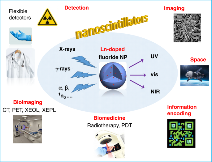
collaborators, describes the world of lanthanide-doped nanoscintillators and photon converters7. The review essentially focuses on lanthanide fluoride nanoparticles doped with various
emissive lanthanide ions. The corresponding nanoscintillators are easy and cheap to process and these materials help solving important problems in medical imaging. One has been mentioned
above, replacing visible-emitting probes with NIR-emitting ones, particularly in the NIR-II biological window (1000–1700 nm). Another one pertains to long-persistent luminescence bioprobes
that are useful for following reactions in cells and, also, for monitoring the advancement of medical therapies8. Yet, most of these probes have to be charged by UV-light before being
introduced into the biological medium or the investigated tissue. In this case, even if the probe emits in the NIR-II range, recharging it in vivo is difficult due to the low penetration of
UV light. On the other hand, X-rays penetrate deeply into tissues and are therefore ideal for recharging these NIR-II emitting nanoprobes. Additionally, nanoscintillators can be introduced
into biocompatible polymers such as polydimethylsiloxane (PDMS) yielding flexible detectors that are convenient for the imaging of curved 3D objects, a difficult, or even impossible, task
with flat-panel X-ray detectors9. A main focus of the review is therefore on X-ray excited optical imaging (XEOL) and X-ray excited persistent luminescence (XEPL). The authors devote a large
section of their manuscript to discussing the mechanisms involved in X-ray to UV, visible, and NIR conversion, as well as those pertaining to the generation of persistent luminescence,
making the review an excellent tutorial for scientists entering the field. Convincing examples of XEOL/XEPL imaging follow suit, including circuit boards, biological material, and various
hidden or encapsulated objects. Comparison between flat-panel detectors and XEPL detection with flexible polymers (sometimes referred to as X-ray luminescence extension imaging, Xr-LEI9) is
particularly convincing. A good share of examples are in the field of biomedicine, with multimodal imaging, radiotherapy monitoring, X-ray enabled photodynamic treatment, to name but a few.
Another important field of application lies in information encoding in luminescent QR codes or for multidimensional information storage. However, all problems associated with the design of
high-performance lanthanide-doped nanoscintillators are not completely solved. In particular combining bright XEOL intensity with fast decay rate is not granted and optimization of the
composition and structure of nanoparticles incorporating several lanthanide ions for broadband detection still needs dedicated efforts. These aspects are presently the subject of intensive
studies and further achievements are on their way. Therefore, perspectives for lanthanide-doped nanoscintillators are bright. Their ease of synthesis and stability are considerable assets
and they definitively enable improving several aspects of crucial applications in industrial imaging, information storage, and biomedicine. Regarding the latter, X-ray excited luminescence
is starting to overtake traditional luminescence optical imaging. Radiation detection is also vital for space applications and for all professionals dealing with radioactive materials. In
particular, flexible detectors incorporated into gloves or textiles (lab coats) could help determining not only the received irradiation dose, but, also, its precise cartography. Moreover,
the ability of 3D imaging could be exploited in diagnosis, for instance in breast mammography. REFERENCES * Nikl, M. & Yoshikawa, A. Recent R&D trends in inorganic single-crystal
scintillator materials for radiation detection. _Adv. Optical Mater._ 3, 463–481 (2015). Article Google Scholar * Jana, A. et al. Perovskite: scintillators, direct detectors, and X-ray
imagers. _Mater. Today_ 55, 110–136 (2022). Article Google Scholar * Liu, F. Z. et al. Halide perovskites and perovskite related materials for particle radiation detection. _Nanoscale_ 14,
6743–6760 (2022). Article Google Scholar * Lu, L. et al. All-inorganic perovskite nanocrystals: next-generation scintillation materials for high-resolution X-ray imaging. _Nanoscale Adv._
4, 680–696 (2022). Article ADS Google Scholar * Bünzli, J. C. G. Lanthanide photonics: shaping the nanoworld. _Trends Chem._ 1, 751–762 (2019). * Zhou, J. J. et al. Impact of lanthanide
nanomaterials on photonic devices and smart applications. _Small_ 14, 1801882 (2018). Article Google Scholar * Lei, L. et al. Next generation lanthanide doped nanoscintillators and photon
converters. _eLight_ 2, 17 (2022). Article Google Scholar * Richard, C. & Viana, B. Persistent X-ray-activated phosphors: mechanisms and applications. _Light Sci. Appl._ 11, 123
(2022). Article ADS Google Scholar * Ou, X. Y. et al. High-resolution X-ray luminescence extension imaging. _Nature_ 590, 410–415 (2021). Article ADS Google Scholar Download references
AUTHOR INFORMATION AUTHORS AND AFFILIATIONS * Institute of Chemical Sciences and Engineering, Swiss Federal Institute of Technology, Lausanne (EPFL), Lausanne, Switzerland Jean-Claude
Georges Bünzli * Department of Biomedical Engineering, Southern University of Science and Technology (SUSTech), Shenzhen, China Jean-Claude Georges Bünzli Authors * Jean-Claude Georges
Bünzli View author publications You can also search for this author inPubMed Google Scholar CORRESPONDING AUTHOR Correspondence to Jean-Claude Georges Bünzli. RIGHTS AND PERMISSIONS OPEN
ACCESS This article is licensed under a Creative Commons Attribution 4.0 International License, which permits use, sharing, adaptation, distribution and reproduction in any medium or format,
as long as you give appropriate credit to the original author(s) and the source, provide a link to the Creative Commons license, and indicate if changes were made. The images or other third
party material in this article are included in the article’s Creative Commons license, unless indicated otherwise in a credit line to the material. If material is not included in the
article’s Creative Commons license and your intended use is not permitted by statutory regulation or exceeds the permitted use, you will need to obtain permission directly from the copyright
holder. To view a copy of this license, visit http://creativecommons.org/licenses/by/4.0/. Reprints and permissions ABOUT THIS ARTICLE CITE THIS ARTICLE Bünzli, JC.G. Lanthanide-doped
nanoscintillators. _Light Sci Appl_ 11, 285 (2022). https://doi.org/10.1038/s41377-022-00987-2 Download citation * Published: 29 September 2022 * DOI:
https://doi.org/10.1038/s41377-022-00987-2 SHARE THIS ARTICLE Anyone you share the following link with will be able to read this content: Get shareable link Sorry, a shareable link is not
currently available for this article. Copy to clipboard Provided by the Springer Nature SharedIt content-sharing initiative