Integrating spatially resolved three-dimensional maldi ims with in vivo magnetic resonance imaging
- Select a language for the TTS:
- UK English Female
- UK English Male
- US English Female
- US English Male
- Australian Female
- Australian Male
- Language selected: (auto detect) - EN
Play all audios:
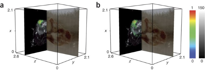
ABSTRACT We have developed a method for integrating three dimensional–volume reconstructions of spatially resolved matrix-assisted laser desorption/ionization imaging mass spectrometry
(MALDI IMS) ion images of whole mouse heads with high-resolution images from other modalities in an animal-specific manner. This approach enabled us to analyze proteomic profiles from MALDI
IMS data with corresponding _in vivo_ data provided by magnetic resonance imaging. Access through your institution Buy or subscribe This is a preview of subscription content, access via your
institution ACCESS OPTIONS Access through your institution Subscribe to this journal Receive 12 print issues and online access $259.00 per year only $21.58 per issue Learn more Buy this
article * Purchase on SpringerLink * Instant access to full article PDF Buy now Prices may be subject to local taxes which are calculated during checkout ADDITIONAL ACCESS OPTIONS: * Log in
* Learn about institutional subscriptions * Read our FAQs * Contact customer support SIMILAR CONTENT BEING VIEWED BY OTHERS TOWARD NANOSCALE MOLECULAR MASS SPECTROMETRY IMAGING VIA
PHYSICALLY CONSTRAINED MACHINE LEARNING ON CO-REGISTERED MULTIMODAL DATA Article Open access 26 June 2020 SPATIAL PROBABILISTIC MAPPING OF METABOLITE ENSEMBLES IN MASS SPECTROMETRY IMAGING
Article Open access 01 April 2023 AUTOMATED ANNOTATION AND VISUALISATION OF HIGH-RESOLUTION SPATIAL PROTEOMIC MASS SPECTROMETRY IMAGING DATA USING HIT-MAP Article Open access 28 May 2021
REFERENCES * Chaurand, P. et al. _Am. J. Pathol._ 165, 1057–1068 (2004). Article CAS Google Scholar * Chaurand, P., Schwartz, S.A. & Caprioli, R.M. _J. Proteome Res._ 3, 245–252
(2004). Article CAS Google Scholar * Khatib-Shahidi, S. et al. _Anal. Chem._ 78, 6448–6456 (2006). Article CAS Google Scholar * Reyzer, M.L. et al. _J. Mass Spectrom._ 38, 1081–1092
(2003). Article CAS Google Scholar * Reyzer, M.L. et al. _Cancer Res._ 64, 9093–9100 (2004). Article CAS Google Scholar * Stoeckli, M. et al. _Nat. Med._ 7, 493–496 (2001). Article
CAS Google Scholar * Crecelius, A.C. et al. _J. Am. Soc. Mass Spectrom._ 16, 1093–1099 (2005). Article CAS Google Scholar * Andersson, M., Groseclose, M.R., Deutch, A.Y. & Caprioli,
R.M. _Nat. Methods_ 5, 101–108 (2008). Article CAS Google Scholar * Paxinos, G. & Franklin, K.B.J. _The Mouse Brain in Stereotaxic Coordinates_. (San Diego, Academic Press, 2001).
Google Scholar * Studholme, C., Hill, D.L.G. & Hawkes, D.J. _Pattern Recognit._ 32, 71–86 (1999). Article Google Scholar * Schwartz, S.A. et al. _Cancer Res._ 65, 7674–7681 (2005).
Article CAS Google Scholar * Atlas, S. _Magnetic Resonance Imaging of the Brain and Spine_. (Philadelphia, Lippincott Williams & Wilkins, 2002). Google Scholar Download references
ACKNOWLEDGEMENTS We would like to thank J. True, R. Baheza and the staff of the Vanderbilt University Institute of Imaging Science Center for Small Animal Imaging for their assistance in
collecting the imaging data presented here. Financial support was provided by the US National Institutes of Health Institute of Biomedical Imaging and Bioengineering, Cancer Institute,
Institute of Neurological Disorders and Stroke and the Institute of General Medical Sciences. AUTHOR INFORMATION AUTHORS AND AFFILIATIONS * Department of Radiology and Radiological Sciences,
Vanderbilt University, Nashville, 37232, 1161 21st Avenue South, Tennessee, USA Tuhin K Sinha, Thomas E Yankeelov & John C Gore * Department of Chemistry, Vanderbilt University,
Nashville, 37232, 1161 21st Avenue South, Tennessee, USA Sheerin Khatib-Shahidi & Richard M Caprioli * Department of Physics and Astronomy, Vanderbilt University, Nashville, 37232, 1161
21st Avenue South, Tennessee, USA Thomas E Yankeelov & John C Gore * Department of Biomedical Engineering, Vanderbilt University, Nashville, 37232, 1161 21st Avenue South, Tennessee, USA
Thomas E Yankeelov & John C Gore * Department of Neurological Surgery, Vanderbilt University, Nashville, 37232, 1161 21st Avenue South, Tennessee, USA Khubaib Mapara & Moneeb
Ehtesham * Department of Cancer Biology, Vanderbilt University, Nashville, 37232, 1161 21st Avenue South, Tennessee, USA Moneeb Ehtesham * Department of Biochemistry, Vanderbilt University,
Nashville, 37232, 1161 21st Avenue South, Tennessee, USA D Shannon Cornett & Richard M Caprioli * Department of Electrical Engineering and Computer Science, Vanderbilt University,
Nashville, 37232, 1161 21st Avenue South, Tennessee, USA Benoit M Dawant * Department of Molecular Physiology and Biophysics, Vanderbilt University, Nashville, 37232, 1161 21st Avenue South,
Tennessee, USA John C Gore Authors * Tuhin K Sinha View author publications You can also search for this author inPubMed Google Scholar * Sheerin Khatib-Shahidi View author publications You
can also search for this author inPubMed Google Scholar * Thomas E Yankeelov View author publications You can also search for this author inPubMed Google Scholar * Khubaib Mapara View
author publications You can also search for this author inPubMed Google Scholar * Moneeb Ehtesham View author publications You can also search for this author inPubMed Google Scholar * D
Shannon Cornett View author publications You can also search for this author inPubMed Google Scholar * Benoit M Dawant View author publications You can also search for this author inPubMed
Google Scholar * Richard M Caprioli View author publications You can also search for this author inPubMed Google Scholar * John C Gore View author publications You can also search for this
author inPubMed Google Scholar CONTRIBUTIONS T.K.S. helped to develop the techniques presented here and assisted in collecting and analyzing the results. S.K.-S. helped to acquire the
blockface and MALDI IMS data. T.E.Y. helped to acquire the _in vivo_ magnetic resonance data. K.M. implanted and provided the mouse with a tumor-laden brain. M.E. provided support and
expertise with the tumor model. D.S.C. provided expertise in collecting the MALDI IMS data. B.M.D. helped to develop accurate coregistration techniques for the MALDI IMS and magnetic
resonance alignment. R.M.C. provided support and expertise for the MALDI IMS data collection. J.C.G. helped to develop the techniques presented here and provided expertise with data analysis
and magnetic resonance data collection. CORRESPONDING AUTHOR Correspondence to Tuhin K Sinha. SUPPLEMENTARY INFORMATION SUPPLEMENTARY TEXT AND FIGURES Supplementary Figures 1–4 and
Supplementary Methods (PDF 1821 kb) SUPPLEMENTARY MOVIE 1 Reconstructed blockface volume of a whole mouse head. (MOV 3915 kb) SUPPLEMENTARY MOVIE 2 Blockface reconstruction results for whole
animals. (MOV 3365 kb) SUPPLEMENTARY MOVIE 3 Coregistered volumetric MALDI IMS data with _in vivo_ magnetic resonance imaging in a tumor-laden mouse brain. (MOV 9343 kb) RIGHTS AND
PERMISSIONS Reprints and permissions ABOUT THIS ARTICLE CITE THIS ARTICLE Sinha, T., Khatib-Shahidi, S., Yankeelov, T. _et al._ Integrating spatially resolved three-dimensional MALDI IMS
with _in vivo_ magnetic resonance imaging. _Nat Methods_ 5, 57–59 (2008). https://doi.org/10.1038/nmeth1147 Download citation * Received: 09 August 2007 * Accepted: 16 November 2007 *
Published: 16 December 2007 * Issue Date: January 2008 * DOI: https://doi.org/10.1038/nmeth1147 SHARE THIS ARTICLE Anyone you share the following link with will be able to read this content:
Get shareable link Sorry, a shareable link is not currently available for this article. Copy to clipboard Provided by the Springer Nature SharedIt content-sharing initiative