Acute postoperative morganella morganii panophthalmitis
- Select a language for the TTS:
- UK English Female
- UK English Male
- US English Female
- US English Male
- Australian Female
- Australian Male
- Language selected: (auto detect) - EN
Play all audios:
MAIN Sir, Endophthalmitis is an uncommon but serious complication of intraocular surgery, often resulting in severe visual loss.1 The endophthalmitis may progress to panophthalmitis if
medical or surgical therapy cannot control the infection. Strains of the _Morganella_ genus are a rare cause of panophthalmitis following trauma and cataract surgery.2 We describe a patient
who developed fulminant _Morganella morganii_ panophthalmitis following vitrectomy with resultant loss of vision. To our knowledge, this is the first reported case of _M. morganii_
panophthalmitis. CASE REPORT A 46-year-old Taiwanese male had sudden loss of vision in right eye for a day due to vitreous haemorrhage. On day 1 after pars plana vitrectomy, the fundus was
visible and the retina was well attached. Unfortunately, his visual acuity decreased to no light perception, and severe ocular pain with eyelid swelling developed rapidly within 2 days after
the operation. He was transferred to our hospital on day 4. On presentation, the patient eyelid appeared to be severely swollen and haemorrhagic chemosis was noted. Mucopurulent pus exuded
from the right eye. Acute postoperative panophthalmitis was diagnosed. Haemogram revealed leukocytosis (16 700/_μ_l) with neutrophils predominant by 80.9%. Pars plana vitrectomy with
anterior chamber irrigation and intravitreal delivery of vancomycin and ceftazidime was performed on day 5. Profuse pus was noted during the operation. Vitreous specimens were obtained for
smear and culture. Intravitreal injections of vancomycin (1 mg/0.1 ml) and ceftazidime (2 mg/0.1 ml) and subconjunctival injections of vancomycin (50 mg/0.5 cm3) and ceftazidime (125 mg/0.5
cm3) were given at the end of the operation. Diffuse retinal whitening and hyperemic disc were noted intraoperatively. The patient's postoperative treatment regimen consisted of: (1)
subconjunctival vancomycin (50 mg/0.5 cm3) and ceftazidime (125 mg/0.5 cm3) every other day (2) intravenous cefazolin (1 g every 8 h) and gentamicin (80 mg every 12 h) and (3) topical
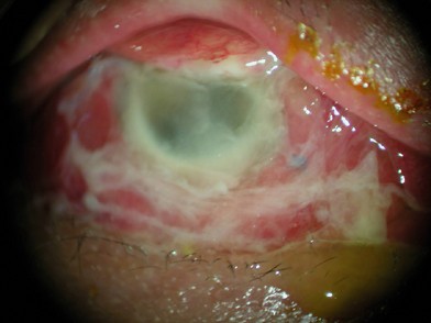
fortified vancomicin (50 mg/cm3) and ceftazidime (50 mg/cm3) every 1 h. However, his eye became more inflamed with mucopurulent discharge, and the ophthalmic examination was difficult to
perform due to severe periorbital swelling (Figure 1). Gram stain of vitreous specimens revealed numerous Gram negative rods (Figure 2), and culture of the anterior chamber aspirates and
vitreous specimens grew many _Morganella morganii_ colonies that were resistant to ampicillin and cefazolin using the disk diffusion method. The extraocular extension was noted by computed
tomography. The systemic antibiotic was then shifted to intravenous ceftriaxone (2 g every 12 h) 1 week after operation. Periorbital swelling with mucopurulent discharge decreased
dramatically after intravenous ceftriaxone use. His eye appeared quiet after a 2-week course of systemic and topical antibiotics with third-generation cephalosporin. Unfortunately, the
patient did not regain any vision despite this aggressive treatment. COMMENT _M. morganii_ is a Gram negative bacillus that belongs to the Enterobacteriaceae family. Within the
Enterobacteriaceae, the genus _Morganella_ is one member of the tribe Proteeae, which includes also the genera _Proteus_ and _Providencia_.3 _M. morganii_ is highly resistant bacillus
susceptible only to _β_-lactamase inhibitors. Strains are often resistant to first-generation cephalosporins.3 _M. morganii_ is a rare but usually devastating cause of postoperative
endophthalmitis.4, 5 In our patient, the clinical picture was fulminating. Even though the early intervention was performed, panopthalmitis with extraocular extension still developed
rapidly. The inflammation did not subside even with aggressive treatment till the third-generation cephalosporin was given intravenously. To our knowledge, this is the first reported case of
_M. morganii_ panophthalmitis. _M morganii_ is a rare aetiologic infectious agent. Very early intensive treatment including systemic antibiotic therapy with the third-generation
cephalosporin is the most important factor in the possible success of avoiding an eye with _M. morganii_ panophthalmitis from evisceration/enucleation. REFERENCES * Nick M, Laura K, Eric B .
Postoperative endophthalmitis. _Curr Opin Ophthalmol_ 2002; 13: 14–18. Article Google Scholar * Irvine WD, Flynn HW, Miller D, Pflugfelded SC . Endophthalmitis caused by gram-negative
organisms. _Arch Ophthalmol_ 1992; 110: 1450–1454. Article CAS Google Scholar * Stock I, Wiedemann B . Identification and natural antibiotic susceptibility of _Morganella morganii_.
_Diagn Microbiol Infect Dis_ 1998; 30: 154–165. Article Google Scholar * Cunningham ET, Whitcher JP, Kim RY . _Morganella morganii_ postoperative endophthalmitis [letter]. _Br J
Ophthalmol_ 1997; 81: 170–171. Article Google Scholar * Tsanaktsidis G, Anarwal SA, Maloof AJ, Chandra J, Mitchell P . Postoperative _Morganella morganii_ endophthalmitis associated with
subclinical urinary tract infection. _J Cataract Refract Surg_ 2003; 29: 1011–1013. Article Google Scholar Download references AUTHOR INFORMATION AUTHORS AND AFFILIATIONS * Department of
Ophthalmology, National Taiwan University Hospital, # 7 Chung-Shan South Rd., Taipei, Taiwan T-J Wang & J-S Huang * Department of Laboratory Medicine and Internal Medicine, National
Taiwan University Hospital, Taipei, Taiwan P-R Hsueh Authors * T-J Wang View author publications You can also search for this author inPubMed Google Scholar * J-S Huang View author
publications You can also search for this author inPubMed Google Scholar * P-R Hsueh View author publications You can also search for this author inPubMed Google Scholar CORRESPONDING AUTHOR
Correspondence to J-S Huang. RIGHTS AND PERMISSIONS Reprints and permissions ABOUT THIS ARTICLE CITE THIS ARTICLE Wang, TJ., Huang, JS. & Hsueh, PR. Acute postoperative _Morganella
morganii_ panophthalmitis. _Eye_ 19, 713–715 (2005). https://doi.org/10.1038/sj.eye.6701613 Download citation * Published: 27 August 2004 * Issue Date: 01 June 2005 * DOI:
https://doi.org/10.1038/sj.eye.6701613 SHARE THIS ARTICLE Anyone you share the following link with will be able to read this content: Get shareable link Sorry, a shareable link is not
currently available for this article. Copy to clipboard Provided by the Springer Nature SharedIt content-sharing initiative