Pin-site myiasis: a rare complication of halo orthosis
- Select a language for the TTS:
- UK English Female
- UK English Male
- US English Female
- US English Male
- Australian Female
- Australian Male
- Language selected: (auto detect) - EN
Play all audios:
ABSTRACT STUDY DESIGN: Case report. OBJECTIVE: To report a rare complication following halo placement for cervical fracture. SETTING: United States University Teaching Hospital. CASE REPORT:
A 39-year-old woman who sustained a spinal cord injury from a C6–7 fracture underwent halo placement. She subsequently developed an infection adjacent to the right posterior pin, which then
became infected with Diptera larvae (maggots), necessitating removal of the pin and debridement of the wound site. CONCLUSION: Halo orthosis continues to be an effective means of
immobilizing the cervical spine. Incidence of complications ranges from 6.4 to 36.0% of cases. Commonly reported complications include pin-site infection, pin penetration, pin loosening,
pressure sores, nerve injury, bleeding, and head ring migration. Pin-site myiasis is rare, with no known reports found in the literature. Poor pin-site care by the patient and her failure to
keep follow-up appointments after development of the initial infection likely contributed to the development of this complication. SIMILAR CONTENT BEING VIEWED BY OTHERS SEVERE COMPLICATION
SUBSEQUENT TO SURGICAL SITE INFECTION AFTER CERVICAL LAMINOPLASTY: A CASE REPORT Article 14 January 2022 FRACTURE-RELATED INFECTION Article 20 October 2022 A NOVEL PRIMARY ANTIBIOTIC
CEMENT-COATED LOCKING PLATE AS A TEMPORARY FIXATION FOR THE TREATMENT OF OPEN TIBIAL FRACTURE Article Open access 11 December 2023 INTRODUCTION Initially described by Perry and Nickel in
1959,1 the halo orthosis continues to be an effective means of stabilizing the cervical spine. Reported incidence of complications associated with halo usage vary from 6.4 to 36.0% of
cases.2, 3, 4 Most complications are associated with pin-site infection, pin penetration through the inner table of the skull, pin loosening, pressure sores, nerve injury, bleeding, and head
ring migration.3, 4, 5 Uncommon infectious complications include the development of intraparenchymal, epidural, or subdural abscesses related to pin penetration.5, 6, 7, 8, 9, 10 In
contrast, pin-site infections are relatively frequent with reported incidence ranging between 5.3 and 20.0% of cases.2, 3, 11, 12, 13 Typically bacterial in origin, most pin-site infections
are effectively treated with pin removal, wound care, and/or antibiotics. We present an unusual case of a pin-site myiasis. Myiasis describes the condition in which a human or other mammal
is infected by Diptera larvae (maggots).14 To our knowledge, this rare complication of halo orthosis has not been previously reported. CASE REPORT A 39-year-old woman with a past medical
history significant for ongoing drug abuse presented as a transfer from an outside facility for management of a gunshot wound to her neck. Initial evaluation at an outside facility was
concerned for a spinal cord injury, prompting institution of high-dose steroids prior to transfer. Upon arrival at our hospital, she was awake and alert but complained of neck pain and
weakness of her right side. On neurologic examination, the patient was noted to have hemiparesis of the right side and decreased left-sided sensation to pin-prick, consistent with
Brown–Sequard syndrome. Evaluation by computed tomography demonstrated an extensive comminuted fracture involving the right C6–7 facet extending to the lamina of C7. Subsequent magnetic
resonance imaging demonstrated increased signal on T2-weighted sequences at the C5–6 level, consistent with spinal cord injury. No significant canal compromise was appreciated, however.
Based on the location and extent of the fracture, a halo orthosis was felt to be a reasonable treatment option. An appropriately sized halo ring was selected. Anteriorly, a pin was placed
approximately 1 cm above the lateral aspect of each eyebrow, while posteriorly, a pin was inserted above either mastoid region. Each pin was then tightened to 8-inch-pounds. The pins were
retightened approximately 24 h later. Routine pin care involving cleaning of the pin sites with a 50% hydrogen peroxide solution every 8 h was instituted. The patient was then transferred to
the rehabilitation service. While on the rehabilitation service, she had significant improvement in her right hemiparesis. Approximately 5 weeks after initial halo placement, a very small
open wound with associated erythema was noted adjacent to the right posterior pin. Cephalexin was started for a presumed infection. After 2 days she was discharged to home. Approximately 2
weeks after discharge, she presented for follow-up to her trauma surgeon who evaluated the area of infection adjacent to her right posterior pin. The previously infected area appeared
unclean and several maggots were reportedly present. The area was debrided and a regimen of levofloxacin was started. After 2 days she presented to the emergency department with the primary
complaint of drainage from her right posterior pin site. Evaluation of the pin site demonstrated a significant degree of erythema with a maggot present within the pin site. Upon removal of
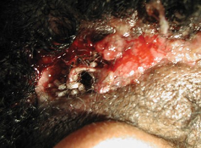
the pin, a large patch of hair and skin adherent to the pin also separated and came away from the surrounding scalp exposing a cavity filled with maggots (Figure 1). The patient subsequently
underwent debridement and irrigation of the scalp. The area was then packed with a Clorpactin-soaked dressing. Two days later, the patient underwent a repeat debridement and irrigation
followed by primary closure. She was then discharged with instructions for a 2-week follow-up for staple removal, but failed to return until 4 weeks later at which point her incision
appeared to be healing without evidence of infection. The patient was subsequently lost to follow-up. DISCUSSION Pin-site infection is one of the most common complications of the halo
orthosis, with one study of 179 patients demonstrating a 20% incidence of infection.3 Subsequent studies, however, have shown a decreased infection rate. Baum _et al_2 found only a 9%
incidence of infection in adults treated with halo immobilization. The decreased infection rate was attributed to several factors including proper application of the halo orthosis with
re-tightening of pins, appropriate patient instruction, and close follow-up. Specifically, the halo orthosis was placed with pins tightened to 6-inch-pounds of torque. Pins were re-torqued
10–15 min and then 24–48 h later. Patient instructions included proper pin-site care including cleaning with hydrogen peroxide twice daily. Follow-up consisted of office visits every 2 weeks
for re-evaluation of the halo orthosis. Similarly, Vertullo _et al_13 found a lower infection rate of 6% in patients who underwent regular re-tightening of the pins to 6–8 inch-pounds at 24
h and then 1 week after halo placement with follow-up evaluation every 2 weeks. Bacterial in origin, treatment of a pin-site infection consists of local wound care and systemic antibiotics.
Removal of the pin with placement at an alternate site may also be performed.2, 3, 11 In this case, the patient underwent proper placement of her halo orthosis including suitable placement
of the pins, tightening of pins to 8 inch-pounds, and re-tightening of pins within 24–48 h. Pin-site care while in rehabilitation was also appropriate, as was the initial management of her
superficial infection with systemic antibiotics. However, the subsequent poor care of her infection and adjacent pin site after discharge, as well as the lack of close follow-up, likely
contributed to the development of myiasis affecting her pin site wound and surrounding scalp. In patients who may be poorly compliant with pin-site care, follow-up visits, or both, the
decision to use a halo orthosis should take into account the likely higher risk for infection. To our knowledge, there have been no previous reports of myiasis involving a halo pin. Maggots
can be classified into obligatory or facultative parasites.15 Obligatory maggots can infect living tissue and are invasive, whereas facultative maggots tend to infect the necrotic tissue of
dead or living hosts. Although uncommon in the United States, wound myiasis has been reported, most often involving the lower extremities of patients with venous stasis ulcers, diabetic
ulcers, pressure ulcers, nonhealing surgical wounds, or traumatic wounds.15 Reports also exist of an infection involving a parietal scalp wound as well as in a shoulder wound.14, 16
Presumably, myiasis occurred in this case via the small infected wound adjacent to the posterior pin. Although the type of maggot, facultative or obligatory, involved in this case is
unclear, the extent of involvement suggests an invasive characteristic. Adequate treatment for myiasis requires the complete removal of the maggots, as was done in this case.16 REFERENCES *
Perry J, Nickel VL . Total cervical spine fusion for neck paralysis. _J Bone Joint Surg Am_ 1959; 41: 37. Article Google Scholar * Baum JA, Hanley EN, Pullekines J . Comparison of halo
complication in adults and children. _Spine_ 1989; 14: 251–252. Article CAS Google Scholar * Garfin SR, Botte MJ, Waters RL, Nickel VL . Complications in the use of the halo fixation
device. _J Bone Joint Surg Am_ 1986; 68: 320–325. Article CAS Google Scholar * Nickel VL, Perry J, Garrett A, Heppenstall M . The halo. A spinal skeletal traction fixation device. _J Bone
Joint Surg Am_ 1968; 50: 1400–1409. Article CAS Google Scholar * Papagelopoulos PJ, Sapkas GS, Kateros KT, Papadakis SA, Vlamis JA, Falagas ME . Halo pin intracranial penetration and
epidural abscess in a patient with a previous cranioplasty: case report and review of the literature. _Spine_ 2001; 26: E463–E467. Article CAS Google Scholar * Celli P, Palatinsky E .
Brain abscess as a complication of cranial traction. _Surg Neurol_ 1985; 23: 594–596. Article CAS Google Scholar * Garfin SR, Botte MJ, Triggs KJ, Nickel VL . Subdural abscess associated
with halo-pin traction. _J Bone Joint Surg Am_ 1988; 70: 1338–1340. Article CAS Google Scholar * Goodman ML, Nelson PB . Brain abscess complicating the use of a halo orthosis.
_Neurosurgery_ 1987; 20: 27–30. Article CAS Google Scholar * Humbyrd DE, Latimer FR, Lonstein JE, Samberg LC . Brain abscess as a complication of halo traction. _Spine_ 1981; 6: 365–368.
Article CAS Google Scholar * Victor DI, Bresnan MJ, Keller RB . Brain abscess complicating the use of halo traction. _J Bone Joint Surg Am_ 1973; 55: 635–639. Article CAS Google Scholar
* Chan RC, Schweigel JF, Thompson GB . Halo-thoracic brace immobilization in 188 patients with acute cervical spine injuries. _J Neurosurg_ 1983; 58: 508–515. Article CAS Google Scholar
* Glaser JA, Whitehill R, Stamp WG, Jane JA . Complications associated with the halo-vest. A review of 245 cases. _J Neurosurg_ 1986; 65: 762–769. Article CAS Google Scholar * Vertullo
CJ, Duke PF, Askin GN . Pin-site complications of the halo thoracic brace with routine pin re-tightening. _Spine_ 1997; 22: 2514–2516. Article CAS Google Scholar * Konkol KA, Longfield
RN, Powers NR, Mehr Z . Wound myiasis caused by _Cochliomyia hominivorax_. _Clin Infect Dis_ 1992; 14: 366. Article CAS Google Scholar * Sherman RA . Wound myiasis in urban and suburban
United States. _Arch Intern Med_ 2000; 160: 2004–2014. Article CAS Google Scholar * Kpea N, Zywocinski C . ‘Flies in the flesh’: a case report and review of cutaneous myiasis. _Cutis_
1995; 55: 47–48. CAS PubMed Google Scholar Download references AUTHOR INFORMATION AUTHORS AND AFFILIATIONS * Department of Neurosurgery, University of Michigan Health System, Ann Arbor,
MI, USA P Park, K R Lodhia, S V Eden & J E McGillicuddy * Spine Center, Tucson Orthopaedic Institute, Tucson, AZ, USA K-U Lewandrowski Authors * P Park View author publications You can
also search for this author inPubMed Google Scholar * K R Lodhia View author publications You can also search for this author inPubMed Google Scholar * S V Eden View author publications You
can also search for this author inPubMed Google Scholar * K-U Lewandrowski View author publications You can also search for this author inPubMed Google Scholar * J E McGillicuddy View author
publications You can also search for this author inPubMed Google Scholar RIGHTS AND PERMISSIONS Reprints and permissions ABOUT THIS ARTICLE CITE THIS ARTICLE Park, P., Lodhia, K., Eden, S.
_et al._ Pin-site myiasis: a rare complication of halo orthosis. _Spinal Cord_ 43, 684–686 (2005). https://doi.org/10.1038/sj.sc.3101773 Download citation * Published: 21 June 2005 * Issue
Date: 01 November 2005 * DOI: https://doi.org/10.1038/sj.sc.3101773 SHARE THIS ARTICLE Anyone you share the following link with will be able to read this content: Get shareable link Sorry, a
shareable link is not currently available for this article. Copy to clipboard Provided by the Springer Nature SharedIt content-sharing initiative KEYWORDS * halo orthosis * infection *
myiasis * maggots