
Intravesicular administration of sodium hyaluronate ameliorates the inflammation and cell proliferation of cystitis cystica et glandularis involving interleukin-6/jak2/stat3 signaling pathway
- Select a language for the TTS:
- UK English Female
- UK English Male
- US English Female
- US English Male
- Australian Female
- Australian Male
- Language selected: (auto detect) - EN
Play all audios:
ABSTRACT Cystitis cystica et glandularis (CCEG) is a chronic cystitis that causes extreme agony in affected patients. However, there are lack of effective conservative treatments. In this
study, it is evident that intravesicular sodium hyaluronate (SH) therapy significantly improved the clinical symptoms of CCEG patients and ameliorated the bladder mucosal inflammation and
cell proliferation characteristics of the disease. Immunohistochemical staining showed that the staining intensities of hyaluronidase (HYAL 1/2), CD44, IL-6 and phosphorylated signal
transducer and activator of transcription 3 (p-Stat3) in bladder mucosal tissue were significantly increased in CCEG patients compared with control patients and that intravesicular SH
treatment suppressed these protein expression. We established a CCEG rat model by treating rats with _E_. _coli_ intravesicularly, and we found that HYAL 1/2 and CD44 expression levels were
significantly increased in the _E_. _coli_ group compared with the NC group. Activation of the IL-6/JAK2/Stat3 pathway and the expression levels of the downstream pro-apoptotic proteins
Mcl-1 and Bcl-xL were also significantly increased in the _E_. _coli_ group compared with the NC group. The above changes were significantly mitigated by intravesicular SH treatment.
Therefore, SH may serve as an effective therapy for CCEG by inhibiting bladder mucosal inflammation and proliferation. SIMILAR CONTENT BEING VIEWED BY OTHERS PLATELET-RICH PLASMA ATTENUATES
THE UPEC-INDUCED CYSTITIS VIA INHIBITING MMP-2,9 ACTIVITIES AND DOWNREGULATION OF NGF AND VEGF IN CANIS LUPUS FAMILIARIS MODEL Article Open access 13 June 2024 4-PBA INHIBITS ENDOPLASMIC
RETICULUM STRESS TO IMPROVE AUTOPHAGIC FLUX IN THE TREATMENT OF PROTAMINE/LIPOPOLYSACCHARIDE-INDUCED INTERSTITIAL CYSTITIS IN RATS Article Open access 28 August 2023 _POTENTILLA CHINENSIS_
AQUEOUS EXTRACT ATTENUATES CYCLOPHOSPHAMIDE-INDUCED HEMORRHAGIC CYSTITIS IN RAT MODEL Article Open access 29 July 2022 INTRODUCTION Cystitis cystica et glandularis (CCEG) is a chronic
reactive inflammatory disorder thought to be attributable to chronic urothelial irritation caused by infections, tumors, calculi or outlet obstructions1. CCEG is characterized by pathologic
proliferative changes in the bladder mucosa. Early studies suggest that CCEG is a precancerous lesion2,3. Recently, more and more evidences suggest that there is no significant causal
relationship between CCEG and bladder malignancies, but there exists phenomenon of coexistence of cystitis glandularis and bladder carcinoma with high ratio4,5,6. The typical symptoms of
CCEG are urinary frequency, urinary urgency, dysuria, and hematuria, which cause extreme discomfort in affected patients and reduce their quality of life. Improvements in cystoscopy and
biopsy techniques have led to a gradual increase in the number of reports about CCEG over the last decade. However, the pathogenesis of CCEG remains unclear7. Given that CCEG is a chronic
reactive inflammatory disorder that causes pathologic proliferative changes in the bladder mucosa, we elected to focus on the mechanisms responsible for the inflammation and cell
proliferation characteristic of CCEG in this study. Increasing amounts of evidence have indicated that inflammatory signals play an important role in sustaining and promoting neoplastic
growth. The pro-inflammatory cytokine IL-6 and its downstream effectors, Janus-activated kinases (JAK2) and signal transducer and activator of transcription 3 (Stat3), have been demonstrated
to play important roles in blocking cell apoptosis and enhancing cell proliferation in various hyperplastic diseases8,9. For example, IL-6 has been reported to induce phosphorylation of
Stat3, which is associated with increases in the expression levels of the anti-apoptotic genes Bcl-xL and Mcl-1 in Barrett’s esophagus, a condition that appears to result from chronic
irritation and is characterized by the replacement of dysplastic squamous epithelial cells with metaplastic intestinal-like columnar epithelial cells10,11. Similar pathologic changes occur
in the bladder in CCEG. The IL-6/JAK2/Stat3 pathway plays a critical role in inflammation and proliferation. Both of inflammation and proliferation occur in the bladder mucosa in CCEG. Thus,
it is hypothesised that the IL-6/JAK2/Stat3 pathway is also involved in CEGG development and progression. Thus, in this study, we investigated the activity of the IL-6/JAK2/Stat3 signaling
pathway and assessed the expression of the downstream anti-apoptotic biomarkers Mcl-1 and Bcl-xL to elucidate the molecular mechanisms underlying the development of CCEG. Glycosaminoglycans
(GAGs) form a thick layer that covers the bladder epithelium to block various irritants, such as chemicals, calculi and bacteria that cause chronic infections12. The protection provided by
GAG layers may prevent the constant evolution of bladder inflammation. GAGs have recently become a novel therapy for the treatment of recurrent urinary tract infections and interstitial
cystitis/painful bladder syndrome (IC/PBS)13,14,15. Endogenous hyaluronic acid (HA) is a key component of GAGs, and recent studies have suggested that intravesicular instillation of sodium
hyaluronate (SH) may promote regeneration of the GAG layers on the bladder urothelium and inhibit IL-6 secretion in bladder tissue16. However, few studies have investigated the effects of SH
treatment on human CCEG. Thus, in the present study, we evaluated the effects of treatment with SH on CCEG patients and elucidated the mechanisms linking the IL-6/JAK2/Stat3 pathway to CCEG
in a CCEG rat model. RESULTS SH AMELIORATED BLADDER MUCOSAL INFLAMMATION AND CELL PROLIFERATION AND THUS IMPROVED THE CLINICAL SYMPTOMS OF CCEG PATIENTS All CCEG patients were given pre-
and post-treatment “PUF Patient Symptom Scale Questionnaires” to assess the effects of treatment with SH on their clinical symptoms. The voiding dairies completed by the patients enrolled
herein were used to estimate their mean daytime urinary frequency and their maximum bladder volume. After SH treatment, CCEG patients displayed significant improvements in their bladder
pain, daytime urinary frequency and maximum bladder volumes (Table 1). With the exception of one patient who experienced two episodes of transient whole-body itching, no patients experienced
severe adverse events during the indicated period. 
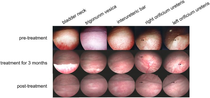
In the control group, the mucosa of the bladder trigonum and bladder neck appeared pink and smooth under 70° cystoscopy. In 16 CCEG
patients, the bladder trigonum and bladder neck displayed white villous, follicular, papillary changes before SH treatment. Eleven of the indicated patients had inflammatory congestion. All
CCEG patients experienced varying degrees of improvement in the appearance of their bladder mucosal tissues after being treated with SH for 6 months, and 6 patients displayed normal bladder
mucosa under cystoscopy, i.e., mucosa that appeared pink and smooth under cystoscopy, after treatment with SH (Fig. 1). HE staining showed that patients in the control group had normal
bladder mucosal tissues with an intact epithelial layer and that no pathological changes had occurred, and no inflammatory cell infiltrates were present in the submucosa. Severe epithelial
damage, Brunn’s nests, cysts and inflammatory cell infiltrates were observed in the submucosa in all the samples obtained from patients in the pre-treatment group. Treatment with SH
significantly ameliorated CCEG-induced inflammation and cell proliferation in the bladder mucosa in the post-treatment group. The patients in this group were found to have an intact
epithelial layer and decreased numbers of Brunn’s nests and cysts and less severe inflammatory cell infiltration compared with their counterparts in the pre-treatment group (Fig. 2). The
data pertaining to patient histological scores, inflammatory cell counts and Brunn’s nest counts demonstrated that CCEG induced severe inflammation and significant cell proliferation in the
pre-treatment group and that these changes were significantly ameliorated in the post-treatment group (Table 1). IMMUNOHISTOCHEMICAL (IHC) ANALYSIS OF HYAL 1/2, CD44, IL-6, P-STAT3, STAT3,
MCL-1 AND BCL-XL IN HUMAN SAMPLES Under pathological conditions, internalized HA is degraded by hyaluronidases (such as HYAL 1/2)17, and the products of this degradation have been
demonstrated to mediate extensive inflammatory responses by interacting with the CD44 receptor18,19,20. We detected the HYAL 1/2 and CD44 expression and localization in human samples using
the semi-quantitative IHC analysis. The results showed that the HYAL 1/2 and CD44 were localized in urothelial cells and their staining intensities were significantly increased in the
pre-treatment group compared with the control group. After SH treatment, the staining intensities of HYAL 1/2 and CD44 were significantly decreased in the post-treatment group (Fig. 3).
Previous researches have shown that endogenous HA fragments induced IL-6 production via a CD44-independent mechanism in bone marrow cells21, and IL-6 activated Stat3 pathway in various
cancers9,22,23. Here, IHC staining was used to examine IL-6 and p-Stat3 expression and localization (Fig. 4). IL-6 and p-Stat3 were localized in urothelial cells and their staining
intensities were significantly increased in the pre-treatment group compared with the control group. After SH treatment, the staining intensities of IL-6 and p-Stat3 were significantly
decreased in the post-treatment group compared with the pre-treatment group. Spearman correlation coefficient test was performed to analyze the relationship between IL-6 and p-Stat3. A
significant positive correlation emerged between the IL-6 H-score and p-Stat3 H-score. (R = 0.655, P = 0.006 in the pre-treatment group; R = 0.546, P = 0.029 in the post-treatment group; and
R = 0.586, P = 0.017 in the H-score difference between the pre-treatment group and the post- treatment group). The total Stat3 expression levels were also compared among pre-treatment,
post-treatment and control group. Figure 4 shows that the biopsy specimens from patients in the pre-treatment, post-treatment and control groups displayed similar levels of total Stat3
expression despite of different levels of p-Stat3 expression, suggesting that increases in p-Stat3 expression are not caused by total Stat3 upregulation and that treatment with SH does not
affect total Stat3 expression. The expression and location of Bcl-xL and Mcl-1 were also detected through IHC staining analysis. Bcl-xL and Mcl-1 were mainly localized in the basal layer of
urothelial cells and their staining intensities were significantly increased in CCEG patients and were significantly decreased after SH treatment (Fig. 5). INTRAVESICULAR ADMINISTRATION OF
SH ATTENUATED _E_. _COLI_-INDUCED CYSTITIS IN RATS The genesis of CCEG has been shown to be strongly correlated with chronic lower urinary tract infections, particularly those caused by _E_.
_coli_ 7. Intravesicular administration of _E_. _coli_ has been reported to induce cystitis glandularis in rats24. Here, we used a similar method to generate an _E_. _coli_-induced CCEG rat
model, whose successful establishment was subsequently confirmed by HE staining. The bladder mucosa in the NC group was normal and consisted of 4-6 layers of epithelial cells of an
identical size. No heterocysts were present, and no inflammatory cellular infiltrates were observed in the submucosal layer. Brunn’s nests, cystitis cystica and cystitis glandularis were
observed in 12 samples in the _E_. _coli_ group. The glands were covered with the transitional epithelium of the urinary tract, and significant inflammatory cell infiltration was noted in
the vicinity of the Brunn’s nests and cysts. Intravesicular administration of HA can improve bacterial cystitis and attenuate cystitis-induced hypercontractility in rats25. Here, we
evaluated the effects of SH in rats with CCEG. The histopathologic changes observed in the _E_. _coli_ + NS group were similar to those observed in the _E_. _coli_ group; however,
inflammatory cell infiltration was less severe in the former group than in the latter group. Moreover, Brunn’s nest and cyst numbers were significantly decreased in the _E_. _coli_ + SH
group compared with the _E_. _coli_ + NS group (Table 2). Additionally, no significant inflammatory cell infiltrates were noted in the vicinity of the Brunn’s nests and cysts in the _E_.
_coli_ + SH group (Fig. 6). SH TREATMENT REDUCED THE EXPRESSION OF HYAL 1/2 AND CD44 IN _E_. _COLI_-INDUCED CYSTITIS GLANDULARIS RATS Real-time quantitative PCR and western blotting were
performed to determine the expression levels of HYAL 1/2 and CD44 in rat bladder mucosal tissues. The results showed that bacterial infection induced increases in the mRNA and protein
expression of HYAL 1/2 and CD44 in the _E_. _coli_ group compared with the NC group. In contrast, their expression levels were significantly decreased in the _E_. _coli_ + SH group compared
with the _E_. _coli_ + NS group, indicating that intravesicular administration of SH plays important roles in decreasing hyaluronidase expression and preventing internalized HA degradation
(Fig. 7a–c and Fig. 8a). IHC staining was performed to detect the expression and location of HYAL1 and CD44, staining showed that HYAL1 mainly located in the cytoplasm of urothelial cells
while CD44 mainly located on their membrane. Their expression trend is consistent with the expression trend of western blot (Fig. 9). SH TREATMENT INHIBITED THE ACTIVATION OF NF-ΚB AND
IL-6/JAK2/STAT3 PATHWAY IN E.COLI-INDUCED CYSTITIS GLANDULARIS RATS NF-κB is not only an important transcription factor in inflammatory responses but also a key player in anti-apoptotic
signaling. We detected the NF-κB p65 activation in rat models by western blotting analysis. Results showed that p-p65 expression levels were significantly increased in the _E_. _coli_ group
compared with the NC group and significantly decreased in the _E_. _coli_ + SH group compared with the _E_. _coli_ + NS group (Fig. 8c and k). We then evaluated the activity of the
IL-6/JAK2/Stat3 pathway and the expression of its downstream anti-apoptotic proteins in rat models using real-time quantitative PCR and western blotting. The results showed that IL-6, p-JAK2
and p-Stat3 expression levels were significantly increased in the _E_. _coli_ group compared with the NC group and significantly decreased in the _E_. _coli_ + SH group compared with the
_E_. _coli_ + NS group (Figs 7 and 8). Moreover, the expression levels of the anti-apoptotic markers, Bcl-xL and Mcl-1, were significantly increased in the _E_. _coli_ group compared with
the NC group and significantly decreased in the _E_. _coli_ + SH group compared with the _E_. _coli_ + NS group (Figs 7 and 8). DISCUSSION CCEG is a common proliferative disorder of the
urinary bladder whose incidence has continued to rise in recent years due to the popularity of cystoscopy26. CCEG patients usually suffer from recurring symptoms of chronic irritation, such
as urinary frequency, urinary urgency, dysuria, suprapubic and perineal discomfort and hematuria27. Most patients who are admitted to the hospital for hematuria or long-term bladder
irritation cannot be cured of their diseases27. The cause of CCEG remains ambiguous; however, the majority of investigators believe that the disease is induced by chronic inflammation or
irritation9,11. Infection causes primary irritation of the urothelium, and long-term chronic infection may cause the transitional epithelium to proliferate into buds, invade the lamina
propria and differentiate into intestinal columnar epithelial mucin-secreting glands (cystitis glandularis) or cystic deposits (cystitis cystica)28. CCEG is diagnosed based on the presence
of characteristic pathological phenomena, such as increases in the numbers of nest-like structures known as Brunn’s nests, secretory cyst development, and lymphocyte infiltration29. In this
study, we established a CCEG rat model via long-term intravesicular instillation of _E_. _coli_ and verified the successful establishment of the model by histopathologically examining tissue
specimens under a microscope after staining them with HE. Currently, treating patients with severe CCEG entails removing the source of irritation; administering antibiotics, NSAIDs, steroid
pulse therapy and anti-allergy drugs; and performing transurethral excision and fulguration of the inflamed area30. Oral medications usually have limited effectiveness, and surgery is too
painful for many patients. Thus, new effective therapies causing only limited amounts of pain are urgently needed for the treatment of CCEG patients. Injury of the GAG layers of the bladder
is a main cause of various types of cystitis. Intravesicular administration of SH, a mucopolysaccharide that constitutes an important proportion of GAGs, has been shown to have excellent
efficacy with respect to the treatment of IC, radiation-induced cystitis or recurrent bacterial cystitis31,32,33,34. However, few studies have focused on the efficacy of intravesicular SH
therapy for CCEG patients. Here, for the first time, we observed the effects of treatment with SH on bladder mucosal inflammation and cell proliferation in CCEG patients. We found that a
course of intravesicular SH treatment significantly attenuated the mucosal inflammation and proliferation characteristic of CCEG in all patients with the disease. Six patients displayed
normal bladder mucosa under cystoscopy, which appeared pink and smooth after SH treatment. Furthermore, HE staining revealed that Brunn’s nest and cyst counts were significantly decreased
and that lymphocytic infiltration in the vicinity of Brunn’s nests and cysts was reduced in the post-treatment group compared with the pre-treatment group. Patients clinical symptoms also
improved—findings supported by our observation of decreases in PUF scores, reductions in daytime urinary frequency and increases in maximum bladder volume in the post-treatment group
compared with the pre-treatment group—and thus exhibited changes consistent with those noted in the experiments in which bladder mucosal histopathology was assessed. As intravesicular SH
therapy improved clinical symptoms and alleviated bladder mucosa proliferation and inflammation in CCEG patients, we investigated the molecular mechanisms underlying the effects of treatment
with SH on CCEG using CCEG animal models. Endogenous HA is a major component of the extracellular matrix under normal conditions, and many studies have shown that HA is degraded into small
fragments by hyaluronidases (such as HYAL1/2) when tissue injury occurs17. HYAL1 degrades HA into oligosaccharides, while HYAL2 degrades HA into intermediate-sized fragments (~20 kDa)35.
CD44 is a major cell surface receptor for small HA fragments, and interactions between CD44 and HA fragments can trigger various downstream signaling pathways, including pathways associated
with inflammation, proliferation, and angiogenesis induction36. In collagen-induced mouse arthritis, small HA fragments produced by HYALs activated CD44 and induced upregulation of the
pro-inflammatory cytokine IL-618. In normal human dermal fibroblasts, low-molecular-weight hyaluronan is believed to activate cells that participate in wound healing by interacting with HA
receptors, such as CD44, and inducing IL-6 expression20. Adding small HA fragments to cultured normal chondrocytes induced severe inflammation by upregulating CD44 expression, activating
NF-κB translocation and upregulating the pro-inflammatory cytokines TNF-α, IL-6 and IL-1β37. In this study, we found that HYAL1/2 and CD44 expression levels were significantly increased in
the bladder mucosa of rats with CCEG compared with the bladder mucosa of normal control rats. These results suggested that endogenous HA was degraded into small fragments by HYAL1/2 and then
interacted with CD44 in the bladder mucosal tissues of rats with CCEG. We concluded that this may be the critical mechanism by which the proliferation and inflammation characteristic of
CCEG occurs. We also postulated that inhibiting endogenous HA degradation or blocking the interactions between small HA fragments and CD44 may prevent the proliferation and inflammation
characteristic of CCEG. In this study, we found that intravesicular SH therapy significantly inhibited HYAL1/2 and CD44 overexpression in CCEG bladder mucosal tissues. Taken together, these
results indicate that intravesicular SH therapy may inhibit the inflammatory process activated by CD44, as well as the initial process through which endogenous HA is degraded. IL-6 is a
critical cytokine in tumorigenesis, and early studies indicated that IL-6 act as a pro-tumorigenic agent in many cancers, implying that it plays an important role in proliferation38,39. IL-6
is also an important marker of acute or chronic inflammation. Previous studies showed that urine IL-6 levels increased in patients with IC/BPS and appeared to be correlated with symptom and
inflammation severity in such patients40,41. Our study showed that IL-6 mRNA and protein expression levels were elevated in CCEG bladder mucosal tissues, suggesting that IL-6 plays a
critical role in the inflammation and proliferation characteristic of CCEG. IL-6/JAK2/Stat3 pathway is a canonical cascade in inflammation and cell proliferation. The elevated activity of
JAK2 and Stat3 is frequently observed in a variety of human malignancies38,42,43. We examined JAK2 and Stat3 activity levels in the indicated tissues and found that p-JAK2 and p-Stat3
expression levels were significantly elevated in the _E_. _coli_ group compared with the NC group. This result was consistent with those of a previous study, which showed that p-Stat3
activation is increased in bladder epithelial cells in CCEG patients. A previous study demonstrated that intravesicular SH therapy significantly decreased cytokine IL-6 secretion, increased
sulfated GAGs production and improved clinical symptoms in patients with IC16,32. Additionally, Stat3 signaling pathway inhibition improved bladder function after urinary bladder
inflammation44. Thus, we speculated that intravesicular SH therapy may also serve as a treatment for CCEG by inhibiting the IL-6/JAK2/Stat3 pathway. We tested this theory in an animal model
and found that IL-6, p-JAK2 and p-Stat3 expression levels were significantly decreased in the bladder mucosal tissues of the _E_. _coli_ + SH group compared with those of the _E_. _coli_
group after the former group received intravesicular SH therapy. The current study demonstrated that intravesicular SH therapy can suppress bladder epithelial cell proliferation and
inflammation through the IL-6/JAK2/Stat3 signaling pathway. After being activated by IL-6, Stat3 dimerizes, translocates to the nucleus, binds to gene-promoter sequences and induces the
expression of specific genes45. Activated Stat3 regulates several genes, including the anti-apoptotic and proliferation-related genes Bcl-xL, Bcl-2, and Mcl-146,47. Bcl-xL and Mcl-1 are
anti-apoptotic members of the Bcl-2 family and are significantly overexpressed in bladder cancer48,49. Therefore, Bcl-xL and Mcl-1 are key regulators of apoptosis in bladder proliferative
diseases. We evaluated Bcl-xL and Mcl-1 expression in the bladder tissues of rats with CCEG. The results showed that Bcl-xL and Mcl-1 expression was significantly increased in the _E_.
_coli_ group compared with the NC group and that intravesicular SH therapy significantly suppressed Bcl-xL and Mcl-1 expression in the corresponding group compared with the _E_. _coli_
group. Given that the changes in Bcl-xL and Mcl-1 expression levels were consistent with the activation of IL-6/JAK2/Stat3 signaling pathway, we speculated that an important mechanism
underlying the upregulation of Mcl-1 and Bcl-xL is the IL-6/JAK2/Stat3 signaling pathway. Previous studies have shown that inducing the expression of the anti-apoptotic proteins in the Bcl-2
family promoted resistance to cisplatin-induced apoptosis by activating the NF-κB pathway in bladder cancer cells and tissues49,50. In this study, the activity of NF-κB p65 were
significantly increased in the _E_. _coli_ group compared with the NC group and significantly decreased after SH treatment. NF-κB is not only an important transcription factor in
inflammatory responses but also a key player in anti-apoptotic signaling. This further confirmed that SH treatment can significantly improve the inflammation and proliferation of bladder
mucosa in CCEG rats. MATERIALS AND METHODS ANTIBODIES AND REAGENTS Antibodies specific for CD44, HYAL-1, HYAL-2, IL-6, Stat3, phospho-Stat3, Mcl-1, Bcl-xL and GAPDH were obtained from Abcam
(Cambridge, UK), antibodies to JAK2, phosphor-JAK2, p65, phosphor-p65 were purchased from Cell Signaling Biotechnology (Hertfordshire, England) and sodium hyaluronate (SH; Cystistat®) was
purchased from Mylan Institutional (Coill Rua, Inverin, County Galway, Republic of Ireland). DIAGNOSIS OF CCEG AND INTRAVESICULAR SH THERAPY Of all the outpatients admitted to the Department
of Urology, Yankuang Group General Hospital, Zoucheng, China, between Mar 2013 and Jun 2016, 16 were histopathologically diagnosed with typical CCEG. The study protocol was approved by the
Institutional Review Board of Yankuang Group General Hospital, Zoucheng, China, and written informed consent was obtained from each patient who participated in the study. The corresponding
author confirmed that all methods were performed in accordance with relevant guidelines and regulations. Following bladder catheterization, each patient received 50 ml of SH solution and
then rested for 1 h. The above treatment was administered once per week for the first 12 weeks of therapy and was then administered once every two weeks for the next 3 months, after which
the participants were followed up for another 3 months. Cystoscopy and biopsy were performed before and after intravesicular SH therapy. EVALUATION OF THE EFFECTIVENESS OF INTRAVESICULAR SH
THERAPY All the patients were given pre- and post-treatment “Pelvic Pain and Urinary/Frequency (PUF) Patient Symptom Scale Questionnaires” to assess the severity of their symptoms. Moreover,
the patients were asked to keep a 3-day voiding diary pre- and post-treatment to record their maximum bladder capacity (MBC) and any episodes of urinary frequency or nocturia. Patient
tissue samples were analyzed by a pathologist blinded to the patients’ clinical characteristics, and bladder histological scores were determined using a four-point scoring system. HUMAN
BLADDER SPECIMEN HARVESTING AND CLASSIFICATION Sixteen bladder tissue specimens were obtained from CCEG patients (age: 42–61 years)—as diagnosed via histopathological analysis—via
cystoscopic tissue biopsy by the Department of Urology, Yankuang Group General Hospital. The above bladder tissue specimens were obtained from the bladder trigonum pre- and post-treatment
and were thus assigned to pre-treatment and post-treatment groups, respectively. Six normal bladder tissue specimens, which were obtained from patients undergoing transurethral bladder tumor
resection, served as normal controls. All patients provided informed consent regarding the use of their samples in the study, which was approved by the Institutional Review Board of
Yankuang Group General Hospital and was performed in accordance with established national and institutional ethical guidelines pertaining to the treatment of human subjects and the use of
human tissue specimens for research. ESTABLISHMENT OF THE _E_. _COLI_–INDUCED CYSTITIS ANIMAL MODEL All animal experiments complied with the ARRIVE guidelines, were performed in accordance
with the National Institutes of Health Guide for the Care and Use of Laboratory Animals (NIH Publications No. 8023, revised 1978) and were approved by the Ethics Committee on the Care and
Use of Laboratory Animals, Qilu Hospital, Shandong University, Jinan, P. R. China. For these experiments, 80 female Sprague–Dawley rats weighing 200–230 g were randomized into blank control
(NC), Escherichia coli (_E_. _coli_), Escherichia coli + saline (_E_. _coli_ + NS) and Escherichia coli + SH (_E_. _coli_ + SH) groups, each of which comprised 20 rats. _E. coli_ DH5-alpha
was chosen and the concentration of _E. coli_ was turbidimetrically determined with the concentration maintained at 5 × 10⁷/ml level. The rats in the NC group were raised under standard
conditions for 2 months and did not receive any treatments. A PE-50 transurethral catheter was inserted into the bladder of each rat in the _E_. _coli_ group to empty the bladder, after
which each rat received an intravesicular infusion of 0.5 mL of _E_. _coli_ and then rested for 30 min. The rats in the indicated group were then treated with _E_. _coli_ three times a week
for 2 months. The rats in the _E_. _coli_ + NS group received the same pre-treatment as the rats in the _E_. _coli_ group and then received intravesicular infusions of NS three times a week
for 2 months. The rats in the _E_. _coli_ + SH group received the same treatment as the rats in the _E_. _coli_ group and then received intravesicular infusions of SH three times a week for
2 months. Within 48 h after the last treatment, all the rats were anesthetized with chloral hydrate solution for bladder tissue specimen harvesting. BLADDER HISTOLOGICAL EVALUATION The
bladder tissue specimens from the patients and each group of rats were fixed in a 4% formaldehyde solution for 48 h, dehydrated in graded ethanol solutions, and then embedded in paraffin
before being cut into 5-μm-thick sections. After staining the specimens with hematoxylin and eosin (HE), we performed morphological analysis of the bladder mucosa by light microscopy. We
examined five random fields in each section at 40 × magnification, after which we calculated a histological score for each section and performed inflammatory cell and Brunn’s nest counts.
The tissue sections were evaluated by a pathologist from the Department of Pathology, Yankuang Group General Hospital, who was blinded to the patients’ and rats’ clinical characteristics.
Bladder histological scores were determined using a four-point scoring system (0, morphologically unremarkable, with no or minimal inflammatory cell inflammation or epithelial changes; 1,
mild inflammatory cell infiltration in the lamina propria characterized by the presence of scattered lymphocytes or monocytes and accompanied by mild chronic edema, hemorrhage or urothelial
changes; 2, moderate inflammatory cell infiltration within the lamina propria that extends into the muscularis propria and is accompanied by moderate chronic edema, hemorrhage, fibrin
deposition or urothelial changes; 3, severe inflammation in the lamina propria and muscularis propria accompanied by urothelial ulceration, severe chronic edema, hemorrhage and fibrin
deposition). REAL-TIME PCR ANALYSIS mRNA expression levels were examined by quantitative real-time PCR (qRT-PCR). Total RNA was extracted from frozen bladder trigonum tissue specimens using
TRIzol reagent (Invitrogen, Carlsbad, CA, USA), according to the manufacturer’s instructions. The total RNA was subsequently reverse transcribed into cDNA with a cDNA Synthesis Kit (TOYOBO,
Shanghai, China). PCR was performed using RealMaster Mix with SYBR Green, which served as a fluorogenic reagent (TOYOBO, Shanghai, China). Each reaction comprised the following steps: 95 °C
for 30 s, followed by 40 cycles of 95 °C for 5 s, 59 °C for 10 s, and 72 °C for 15 s. The expression level of each target gene was measured in triplicate in at least three independent
experiments. The relative mRNA expression levels of the indicated genes were calculated using the ΔΔCT method and normalized to those of GAPDH, which was used as an endogenous control. The
sequences of all the primers used for this experiment are listed in Table 3. IMMUNOBLOTTING Total protein was extracted from the frozen bladder trigonum tissue specimens using lysis buffer
(Beyotime, Shanghai, China), and the protein concentrations were determined using a BCA Assay Kit (Beyotime, Shanghai, China), according to the manufacturer’s instructions. The proteins
(20–80 µg) were separated via SDS-PAGE and transferred onto PVDF membranes using a wet transfer apparatus (Bio-Rad, Hercules, CA, USA). The membranes were then blocked in 5% non-fat milk in
TBST (0.1% Tween20 in Tris-buffered saline) before being incubated with anti-HYAL-1 (1:1000), anti-HYAL-2 (1:1000), anti-CD44 (diluted 1:1000), anti-IL6 (1:1000), anti-p-Stat3 (1:1000),
anti-Stat3 (1:1000), anti-p-JAK2 (1:1000), anti-JAK2 (1:1000), anti-p-p65 (1:1000), anti-p65 (1:1000), anti-Bcl-xL (1:1000), anti-Mcl-1 (1:1000), and anti-GAPDH (1:1000) antibodies overnight
at 4 °C. The membranes were subsequently incubated with the appropriate secondary antibodies (1:5000) for 2 h at room temperature, after which the protein bands were visualized by enhanced
chemiluminescence (Millipore, Massachusetts, USA) and detected using an ImageQuant LAS4000 Mini Chemiluminescence Reader (GE, Fairfield, Connecticut USA). Protein expression was analyzed
with ImageJ software. IMMUNOHISTOCHEMICAL ANALYSIS Five-micrometer-thick serial sections of bladder mucosal tissues were incubated with the appropriate primary antibodies overnight at 4 °C
before being incubated with the appropriate secondary antibodies for 25 min at room temperature. The sections were then treated with peroxidase-marked streptavidin/peroxidase before being
examined under an Olympus microscope (model BX-51, Japan). We used a semi-quantitative scoring system51 to grade the intensity of the immunoreactions. The positively stained cells in each
bladder specimen were scored according to their staining intensity, which was graded using the following scale: 0 (no staining), +1 (weak but detectable staining), +2 (moderate staining) and
+3 (intense staining). Five areas in each slide were evaluated under a microscope at a magnification of 40×. We calculated the H-score of each tissue sample by multiplying the percentage of
cells in each intensity category by the corresponding staining intensity score and then adding these products together. The calculation was performed using the following formula: H-score =
∑(Pc × s), where s represents the intensity score, and Pc is the corresponding cell percentage. STATISTICAL ANALYSIS All data are presented as the mean ± standard deviation unless otherwise
stated. Statistical analysis was performed by a blinded investigator using SPSS 19.0. The data were analyzed with Student’s tests. Associations with IL-6 and p-Stat3 were tested using
nonparametric tests (Spearman correlation coefficient). P < 0.05 was considered statistically significant. All experiments were repeated independently at least three times. DATA AVAILABLE
STATEMENTS The datasets generated and analysed during the current study are not publicly available due to personal health information and exposure of his private parts, but are available
from the corresponding author on reasonable request. CONCLUSION This clinical study showed that intravesicular SH treatment had a significant effect on CCEG patients and that the
IL-6/JAK2/Stat3 pathway is involved in the pathogenesis of CCEG. The animal experimental findings showed that treatment with SH decreased hyaluronidase expression, inhibited endogenous HA
degradation, and reduced the interactions between HA degradation products and CD44 and thus suppressed CD44-dependent IL-6/JAK2/Stat3 pathway activation and downstream anti-apoptotic protein
expression, namely, Mcl-1 and Bcl-xL expression. Therefore, intravesicular SH treatment may serve as an effective therapy for CCEG by inhibiting inflammation and proliferation. REFERENCES *
Wong-You-Cheong, J. J., Woodward, P. J., Manning, M. A. & Davis, C. J. From the archives of the AFIP: Inflammatory and nonneoplastic bladder masses: radiologic-pathologic correlation.
_Radiographics: a review publication of the Radiological Society of North America, Inc_ 26, 1847–1868, https://doi.org/10.1148/rg.266065126 (2006). Article Google Scholar * Garcia Rojo, D.
_et al_. [Transformation of glandular cystitis into bladder transitional carcinoma with adenocarcinoma areas]. _Archivos espanoles de urologia_ 50, 187–189 (1997). CAS PubMed Google
Scholar * Medina Perez, M., Valero Puerta, J. A., Valpuesta Fernandez, I. & Sanchez Gonzalez, M. [Bladder adenocarcinoma with glandular cystitis areas]. _Archivos espanoles de urologia_
54, 254–256 (2001). CAS PubMed Google Scholar * Smith, A. K., Hansel, D. E. & Jones, J. S. Role of cystitis cystica et glandularis and intestinal metaplasia in development of bladder
carcinoma. _Urology_ 71, 915–918, https://doi.org/10.1016/j.urology.2007.11.079 (2008). Article PubMed Google Scholar * Yi, X. _et al_. Cystitis glandularis: A controversial premalignant
lesion. _Oncology letters_ 8, 1662–1664, https://doi.org/10.3892/ol.2014.2360 (2014). PubMed PubMed Central Google Scholar * Gordetsky, J. & Epstein, J. I. Intestinal metaplasia of
the bladder with dysplasia: a risk factor for carcinoma? _Histopathology_ 67, 325–330, https://doi.org/10.1111/his.12661 (2015). Article PubMed Google Scholar * Chen, Z., Ye, Z. &
Zeng, W. Clinical investigation on the correlation between lower urinary tract infection and cystitis glandularis. _Journal of Huazhong University of Science and Technology_. _Medical
sciences = Hua zhong ke ji da xue xue bao. Yi xue Ying De wen ban=Huazhong keji daxue xuebao. Yixue Yingdewen ban_ 24, 303–304 (2004). Article CAS Google Scholar * Xu, Y. Y. _et al_.
Regulation of CD44v6 expression in gastric carcinoma by the IL-6/STAT3 signaling pathway and its clinical significance. _Oncotarget_. https://doi.org/10.18632/oncotarget.17435 (2017). Google
Scholar * Culig, Z. & Puhr, M. Interleukin-6 and prostate cancer: Current developments and unsolved questions. _Molecular and cellular endocrinology_.
https://doi.org/10.1016/j.mce.2017.03.012 (2017). PubMed Google Scholar * Dvorakova, K. _et al_. Increased expression and secretion of interleukin-6 in patients with Barrett’s esophagus.
_Clinical cancer research: an official journal of the American Association for Cancer Research_ 10, 2020–2028 (2004). Article CAS Google Scholar * Flejou, J. F. Histological assessment of
oesophageal columnar mucosa. Best practice & research. _Clinical gastroenterology_ 22, 671–686, https://doi.org/10.1016/j.bpg.2008.03.003 (2008). PubMed Google Scholar * Klingler, C.
H. Glycosaminoglycans: how much do we know about their role in the bladder. _Urologia_ 83(Suppl 1), 11–14, https://doi.org/10.5301/uro.5000184 (2016). Article PubMed Google Scholar *
Cervigni, M. Interstitial cystitis/bladder pain syndrome and glycosaminoglycans replacement therapy. _Translational andrology and urology_ 4, 638–642,
https://doi.org/10.3978/j.issn.2223-4683.2015.11.04 (2015). PubMed PubMed Central Google Scholar * Fiander, N. Painful bladder syndrome and interstitial cystitis: treatment options.
_British journal of nursing_ 22(S26), S28–33 (2013). Google Scholar * Lazzeri, M. _et al_. Managing chronic bladder diseases with the administration of exogenous glycosaminoglycans: an
update on the evidence. _Therapeutic advances in urology_ 8, 91–99, https://doi.org/10.1177/1756287215621234 (2016). Article CAS PubMed Google Scholar * Rooney, P., Srivastava, A.,
Watson, L., Quinlan, L. R. & Pandit, A. Hyaluronic acid decreases IL-6 and IL-8 secretion and permeability in an inflammatory model of interstitial cystitis. _Acta biomaterialia_ 19,
66–75, https://doi.org/10.1016/j.actbio.2015.02.030 (2015). Article CAS PubMed Google Scholar * Misra, S., Hascall, V. C., Markwald, R. R. & Ghatak, S. Interactions between
Hyaluronan and Its Receptors (CD44, RHAMM) Regulate the Activities of Inflammation and Cancer. _Frontiers in immunology_ 6, 201, https://doi.org/10.3389/fimmu.2015.00201 (2015). Article
PubMed PubMed Central CAS Google Scholar * Campo, G. M. _et al_. The inhibition of hyaluronan degradation reduced pro-inflammatory cytokines in mouse synovial fibroblasts subjected to
collagen-induced arthritis. _Journal of cellular biochemistry_ 113, 1852–1867, https://doi.org/10.1002/jcb.24054 (2012). Article CAS PubMed Google Scholar * Oldenburg, D. _et al_. CD44
and RHAMM are essential for rapid growth of bladder cancer driven by loss of Glycogen Debranching Enzyme (AGL). _BMC cancer_ 16, 713, https://doi.org/10.1186/s12885-016-2756-5 (2016).
Article PubMed PubMed Central Google Scholar * Vistejnova, L. _et al_. Low molecular weight hyaluronan mediated CD44 dependent induction of IL-6 and chemokines in human dermal
fibroblasts potentiates innate immune response. _Cytokine_ 70, 97–103, https://doi.org/10.1016/j.cyto.2014.07.006 (2014). Article CAS PubMed Google Scholar * Vincent, T., Jourdan, M.,
Sy, M. S., Klein, B. & Mechti, N. Hyaluronic acid induces survival and proliferation of human myeloma cells through an interleukin-6-mediated pathway involving the phosphorylation of
retinoblastoma protein. _The Journal of biological chemistry_ 276, 14728–14736, https://doi.org/10.1074/jbc.M003965200 (2001). Article CAS PubMed Google Scholar * Hu, B. _et al_. HIC1
attenuates invasion and metastasis by inhibiting the IL-6/STAT3 signalling pathway in human pancreatic cancer. _Cancer letters_ 376, 387–398, https://doi.org/10.1016/j.canlet.2016.04.013
(2016). Article CAS PubMed Google Scholar * Quan, Z. _et al_. Interleukin 6 induces cell proliferation of clear cell renal cell carcinoma by suppressing hepaCAM via the STAT3-dependent
up-regulation of DNMT1 or DNMT3b. _Cellular signalling_ 32, 48–58, https://doi.org/10.1016/j.cellsig.2017.01.017 (2017). Article CAS PubMed Google Scholar * Liu, X., Chen, Z. & Ye,
Z. Etiological study on cystitis glandularis caused by bacterial infection. _Journal of Huazhong University of Science and Technology_. _Medical sciences = Hua zhong ke ji da xue xue bao. Yi
xue Ying De wen ban = Huazhong keji daxue xuebao. Yixue Yingdewen ban_ 27, 678–680, https://doi.org/10.1007/s11596-007-0615-y (2007). Article Google Scholar * Yildiz, N. _et al_.
Intravesical hyaluronic acid treatment improves bacterial cystitis and reduces cystitis-induced hypercontractility in rats. _International journal of urology: official journal of the
Japanese Urological Association_ 22, 598–603, https://doi.org/10.1111/iju.12752 (2015). Article CAS Google Scholar * Lin, H. Y. _et al_. Cystitis glandularis mimics bladder cancer–three
case reports and literature review. _The Kaohsiung journal of medical sciences_ 17, 102–106 (2001). CAS PubMed Google Scholar * Li, A. _et al_. Clinical character of cystitis glandularis
accompanied with upper urinary tract obstruction. _Canadian Urological Association journal = Journal de l’Association des urologues du Canada_ 7, E708–710, https://doi.org/10.5489/cuaj.359
(2013). Article PubMed PubMed Central Google Scholar * Riaz, A., Casalino, D. D. & Dalton, D. P. Cystitis cystica and cystitis glandularis causing ureteral obstruction. _The Journal
of urology_ 187, 1059–1060, https://doi.org/10.1016/j.juro.2011.12.021 (2012). Article PubMed Google Scholar * Noda, S. & Eto, K. Histopathological studies on the cystic formation of
the human urothelium. _The Kurume medical journal_ 37, 55–65 (1990). Article CAS PubMed Google Scholar * Takizawa, N. _et al_. Novel strategy for cystitis glandularis: Oral treatment
with cyclooxygenase-2 inhibitor. _International journal of urology: official journal of the Japanese Urological Association_ 23, 706–708, https://doi.org/10.1111/iju.13121 (2016). Article
CAS Google Scholar * Raymond, I. _et al_. The clinical effectiveness of intravesical sodium hyaluronate (cystistat(R)) in patients with interstitial cystitis/painful bladder syndrome and
recurrent urinary tract infections. _Current urology_ 6, 93–98, https://doi.org/10.1159/000343517 (2012). Article PubMed PubMed Central Google Scholar * Engelhardt, P. F., Morakis, N.,
Daha, L. K., Esterbauer, B. & Riedl, C. R. Long-term results of intravesical hyaluronan therapy in bladder pain syndrome/interstitial cystitis. _International urogynecology journal_ 22,
401–405, https://doi.org/10.1007/s00192-010-1294-y (2011). Article PubMed Google Scholar * Shao, Y., Lu, G. L. & Shen, Z. J. Comparison of intravesical hyaluronic acid instillation
and hyperbaric oxygen in the treatment of radiation-induced hemorrhagic cystitis. _BJU international_ 109, 691–694, https://doi.org/10.1111/j.1464-410X.2011.10550.x (2012). Article PubMed
Google Scholar * Torella, M. _et al_. Intravesical therapy in recurrent cystitis: a multi-center experience. _Journal of infection and chemotherapy: official journal of the Japan Society of
Chemotherapy_ 19, 920–925, https://doi.org/10.1007/s10156-013-0609-6 (2013). Article CAS Google Scholar * Wang, F. _et al_. HYAL1 and HYAL2 inhibit tumour growth _in vivo_ but not _in
vitro_. _PloS one_ 3, e3031, https://doi.org/10.1371/journal.pone.0003031 (2008). Article PubMed PubMed Central ADS CAS Google Scholar * Misra, S. _et al_. Hyaluronan-CD44 interactions
as potential targets for cancer therapy. _The FEBS journal_ 278, 1429–1443, https://doi.org/10.1111/j.1742-4658.2011.08071.x (2011). Article CAS PubMed PubMed Central Google Scholar *
Campo, G. M. _et al_. Small hyaluronan oligosaccharides induce inflammation by engaging both toll-like-4 and CD44 receptors in human chondrocytes. _Biochemical pharmacology_ 80, 480–490,
https://doi.org/10.1016/j.bcp.2010.04.024 (2010). Article CAS PubMed Google Scholar * Grivennikov, S. _et al_. IL-6 and Stat3 are required for survival of intestinal epithelial cells and
development of colitis-associated cancer. _Cancer cell_ 15, 103–113, https://doi.org/10.1016/j.ccr.2009.01.001 (2009). Article CAS PubMed PubMed Central Google Scholar * Hu, B. _et
al_. Microbiota-induced activation of epithelial IL-6 signaling links inflammasome-driven inflammation with transmissible cancer. _Proceedings of the National Academy of Sciences of the
United States of America_ 110, 9862–9867, https://doi.org/10.1073/pnas.1307575110 (2013). Article CAS PubMed PubMed Central ADS Google Scholar * Erickson, D. R. _et al_. A comparison
of multiple urine markers for interstitial cystitis. _The Journal of urology_ 167, 2461–2469 (2002). Article CAS PubMed Google Scholar * Daha, L. K., Lazar, D., Simak, R. & Pfluger,
H. Is there a relation between urinary interleukin-6 levels and symptoms before and after intra-vesical glycosaminoglycan substitution therapy in patients with bladder pain
syndrome/interstitial cystitis? _International urogynecology journal and pelvic floor dysfunction_ 18, 1449–1452, https://doi.org/10.1007/s00192-007-0354-4 (2007). Article PubMed Google
Scholar * Lesina, M. _et al_. Stat3/Socs3 activation by IL-6 transsignaling promotes progression of pancreatic intraepithelial neoplasia and development of pancreatic cancer. _Cancer cell_
19, 456–469, https://doi.org/10.1016/j.ccr.2011.03.009 (2011). Article CAS PubMed Google Scholar * Bromberg, J. & Wang, T. C. Inflammation and cancer: IL-6 and STAT3 complete the
link. _Cancer cell_ 15, 79–80, https://doi.org/10.1016/j.ccr.2009.01.009 (2009). Article CAS PubMed PubMed Central Google Scholar * Cheppudira, B. P. _et al_. Involvement of JAK-STAT
signaling/function after cyclophosphamide-induced bladder inflammation in female rats. _American journal of physiology. Renal physiology_ 297, F1038–1044,
https://doi.org/10.1152/ajprenal.00110.2009 (2009). Article CAS PubMed PubMed Central Google Scholar * Fursov, N., Gates, I. V., Panavas, T., Giles-Komar, J. & Powers, G.
Development and utilization of activated STAT3 detection assays for screening a library of secreted proteins. _Assay and drug development technologies_ 9, 420–429,
https://doi.org/10.1089/adt.2010.0348 (2011). Article CAS PubMed Google Scholar * Yue, P. _et al_. Hydroxamic Acid and Benzoic Acid-Based STAT3 Inhibitors Suppress Human Glioma and
Breast Cancer Phenotypes _In Vitro_ and _In Vivo_. _Cancer research_ 76, 652–663, https://doi.org/10.1158/0008-5472.CAN-14-3558 (2016). Article CAS PubMed Google Scholar * Banerjee, K.
& Resat, H. Constitutive activation of STAT3 in breast cancer cells: A review. _International journal of cancer_ 138, 2570–2578, https://doi.org/10.1002/ijc.29923 (2016). Article CAS
PubMed Google Scholar * Kunze, D., Erdmann, K., Froehner, M., Wirth, M. P. & Fuessel, S. Enhanced inhibition of bladder cancer cell growth by simultaneous knockdown of anti-apoptotic
Bcl-xL and survivin in combination with chemotherapy. _International journal of molecular sciences_ 14, 12297–12312, https://doi.org/10.3390/ijms140612297 (2013). Article PubMed PubMed
Central CAS Google Scholar * Wu, M. _et al_. URGCP/URG4 promotes apoptotic resistance in bladder cancer cells by activating NF-kappaB signaling. _Oncotarget_ 6, 30887–30901,
https://doi.org/10.18632/oncotarget.5134 (2015). Article PubMed PubMed Central Google Scholar * Tan, S. T., Liu, S. Y. & Wu, B. TRIM29 Overexpression Promotes Proliferation and
Survival of Bladder Cancer Cells through NF-kappaB Signaling. _Cancer research and treatment: official journal of Korean Cancer Association_ 48, 1302–1312,
https://doi.org/10.4143/crt.2015.381 (2016). Article Google Scholar * Zhao, S. H. _et al_. Effect of ouabain on myocardial ultrastructure and cytoskeleton during the development of
ventricular hypertrophy. _Heart and vessels_ 28, 101–113, https://doi.org/10.1007/s00380-011-0219-0 (2013). Article PubMed Google Scholar Download references ACKNOWLEDGEMENTS This study
was supported by grants from the Natural Science Foundation of China 81600220; Shandong Provincial Natural Science Foundation BS2014YY020; Major Scientific Projects of Yankuang Group
YK2015A017. AUTHOR INFORMATION Author notes * Yongliang Ni and Shaohua Zhao contributed equally to this work. AUTHORS AND AFFILIATIONS * Department of Urology, Shandong University Qilu
Hospital, Jinan, Shandong, 250012, China Yongliang Ni & Benkang Shi * Department of Geriatrics, Shandong University Qilu Hospital, Jinan, Shandong, 250012, China Shaohua Zhao *
Department of Traditional Chinese Medicine, Yankuang Group General Hospital, Zoucheng, Shandong, 273500, China Xiaoxuan Yin * Department of Urology, Yankuang Group General Hospital,
Zoucheng, Shandong, 273500, China Yongliang Ni, Haixin Wang & Shoubin Jiao * Department of Pathology, Yankuang Group General Hospital, Zoucheng, Shandong, 273500, China Qianqian Guang,
Guangxia Hu & Yi Yang Authors * Yongliang Ni View author publications You can also search for this author inPubMed Google Scholar * Shaohua Zhao View author publications You can also
search for this author inPubMed Google Scholar * Xiaoxuan Yin View author publications You can also search for this author inPubMed Google Scholar * Haixin Wang View author publications You
can also search for this author inPubMed Google Scholar * Qianqian Guang View author publications You can also search for this author inPubMed Google Scholar * Guangxia Hu View author
publications You can also search for this author inPubMed Google Scholar * Yi Yang View author publications You can also search for this author inPubMed Google Scholar * Shoubin Jiao View
author publications You can also search for this author inPubMed Google Scholar * Benkang Shi View author publications You can also search for this author inPubMed Google Scholar
CONTRIBUTIONS Y.L.N. and S.H.Z. designed the research; Y.L.N., X.X.Y., H.X.W., Q.Q.G. and G.X.H. conducted the studies; Y.Y. and S.B.J. analyzed the data and prepared the manuscript; B.K.S.
and S.H.Z. guided the experiments and edited the paper. All authors read and approved the manuscript. CORRESPONDING AUTHOR Correspondence to Benkang Shi. ETHICS DECLARATIONS COMPETING
INTERESTS The authors declare that they have no competing interests. ADDITIONAL INFORMATION PUBLISHER'S NOTE: Springer Nature remains neutral with regard to jurisdictional claims in
published maps and institutional affiliations. RIGHTS AND PERMISSIONS OPEN ACCESS This article is licensed under a Creative Commons Attribution 4.0 International License, which permits use,
sharing, adaptation, distribution and reproduction in any medium or format, as long as you give appropriate credit to the original author(s) and the source, provide a link to the Creative
Commons license, and indicate if changes were made. The images or other third party material in this article are included in the article’s Creative Commons license, unless indicated
otherwise in a credit line to the material. If material is not included in the article’s Creative Commons license and your intended use is not permitted by statutory regulation or exceeds
the permitted use, you will need to obtain permission directly from the copyright holder. To view a copy of this license, visit http://creativecommons.org/licenses/by/4.0/. Reprints and
permissions ABOUT THIS ARTICLE CITE THIS ARTICLE Ni, Y., Zhao, S., Yin, X. _et al._ Intravesicular administration of sodium hyaluronate ameliorates the inflammation and cell proliferation of
cystitis cystica et glandularis involving interleukin-6/JAK2/Stat3 signaling pathway. _Sci Rep_ 7, 15892 (2017). https://doi.org/10.1038/s41598-017-16088-9 Download citation * Received: 16
June 2017 * Accepted: 06 November 2017 * Published: 21 November 2017 * DOI: https://doi.org/10.1038/s41598-017-16088-9 SHARE THIS ARTICLE Anyone you share the following link with will be
able to read this content: Get shareable link Sorry, a shareable link is not currently available for this article. Copy to clipboard Provided by the Springer Nature SharedIt content-sharing
initiative